
Fig. 6
- ID
- ZDB-FIG-250916-43
- Publication
- Muir et al., 2025 - A subset of neutrophil phagosomes is characterised by pulses of Class I PI3K activity
- Other Figures
- All Figure Page
- Back to All Figure Page
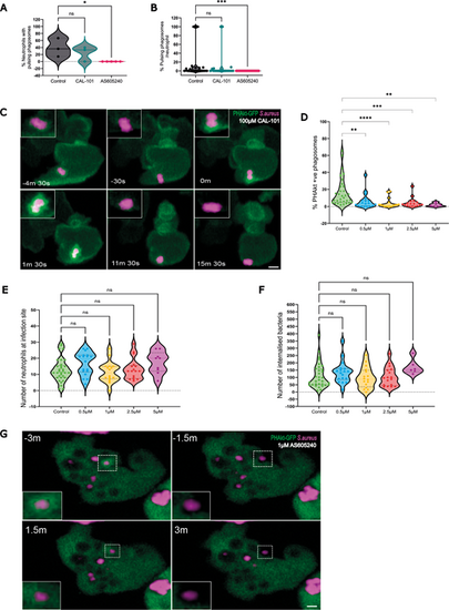
Pulses of PHAkt occur despite inhibition with CAL-101 and are abolished by AS605240. (A) Violin plot showing the percentage of neutrophils that have pulsing phagosomes. Timelapse started 2 h post infection following 30 min incubation of Tg(lyz:PHAkt-EGFP)i277 larvae with 100 µm CAL-101 and 1 µm AS605240. Data shown are the median with the 25th and 75th percentiles from 87 neutrophils, 11 independent larvae, 11 experiments. (B) Violin plot showing the percentage of pulsing phagosomes in each neutrophil in a timelapse starting 2 h post infection following 30 min incubation of Tg(lyz:PHAkt-eGFP)i277 larvae with 100 µm CAL-101 and 1 µm AS605240. Data shown are the median with the 25th and 75th percentiles from 780 phagosomes, 11 independent larvae, 11 experiments. (C) Sequential images illustrating pulsatile recruitment of PHAkt-eGFP to a phagosome exposed to 100 µm CAL-101. Scale bar: 2 µm. (D) Violin plot showing quantification of the percentage of PHAkt-eGFP+ phagosomes 2 h post infection following 30 min incubation with AS605240. Data shown are the median with the 25th and 75th percentiles from six independent larvae, from six experiments. (E) Violin plot showing quantification of the number of neutrophils at a S. aureus infection site 2 h post infection following 30 min incubation with AS605240. Data shown are the median with the 25th and 75th percentiles from six independent larvae, six experiments. (F) Violin plot showing quantification of the number of bacteria/neutrophils 2 h post infection following 30 min incubation with AS605240. Data shown are the median with the 25th and 75th percentiles from six independent larvae, six experiments. (G) Sequential images illustrating that PHAkt-eGFP recruits to neutrophil phagosomes prior to exposure to 1 µm AS605240 (3 min before attempted re-phagocytosis (−3 min). Bacteria are then released from the phagosome (−1.5 min). Following exposure to 1 µm AS605240, the neutrophil attempts to re-phagocytose the bacteria, but PHAkt-eGFP does not recruit to the phagosome (1.5 min), and the bacteria are released from the phagosome (3 min). Scale bar: 2 µm. *P<0.05, **P<0.005, ***P<0.001, ****P<0.0001 (Kruskal–Wallis with multiple comparisons). |