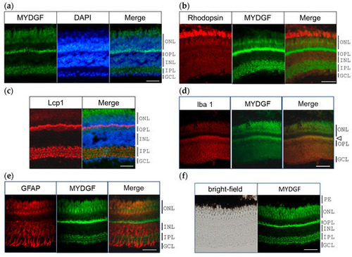
MYDGF-producing cells in the zebrafish retina 1 h after ONI. (a) Double fluorescence staining of MYDGF (green) and nuclear staining with DAPI (blue). The MYDGF-positive cells included photoreceptors in the ONL, the border of the ONL and OPL, and the cells in the IPL (Figure 2a). (b) Fluorescent double staining of the photoreceptor marker proteins rhodopsin (red) and MYDGF (green). (c) Triple fluorescence staining of MYDGF (green), nuclear staining with DAPI (blue), and leukocyte marker Lcp1 (red). Lcp1 clearly colocalized with the MYDGF-positive distribution at the ONL-OPL border regions and in the IPL. (d) The microglial marker protein Iba1 colocalized with the MYDGF-positive region at the ONL-OPL border regions (indicated by the arrowhead). (e) GFAP-positive Müller cells showed almost no co-localization with MYDGF. (f) The pigment epithelium (PE), observed as black under brightfield microscopy, was found to lack MYDGF production. Representative images from 3–4 independent experiments of IHC are shown. Scale bar = 50 μm.
|
