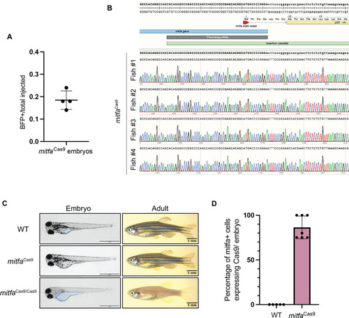
Figure 1—figure supplement 1.

Validation of Cas9 knock-in.

(A) Integration efficiency of mitfa-targeted P2A-Cas9, gamma-cry:BFP vector. n=4 independent experiments are shown with 50 embryos screened for each experiment. BFP+ eyes were detected in a total of 37/200 embryos. Error bars, SD. (B) Sanger sequencing results from four F1 fish with BFP+ eyes. Primers were designed with the forward primer located in the genomic mitfa locus and reverse primer within the Cas9 insertion cassette so that amplification can only occur if there is integration in the correct orientation. SnapGene was used to align sequences. (C) Images of 3-day-old and 3-month-old clutchmates from a mitfaCas9 in-cross. (D) Quantification of the percentage of mitfa-positive cells that have Cas9 expression in each embryo. The presence of mitfa and Cas9 RNA was assessed by confocal microscopy from fluorescence in situ hybridization (FISH) chain reaction on 3 days post-fertilization (dpf) wild-type and mitfaCas9 embryos. Out of 28 mitfa+ cells assessed across n=7 embryos, 24 had detectable Cas9 RNA.

Expression Data

Expression Detail
Antibody Labeling
Phenotype Data

Phenotype Detail
Acknowledgments
This image is the copyrighted work of the attributed author or publisher, and ZFIN has permission only to display this image to its users. Additional permissions should be obtained from the applicable author or publisher of the image. Full text @ Elife