
Fig. 4
- ID
- ZDB-FIG-250828-15
- Publication
- Huang et al., 2025 - Sox11 genes affect neuronal differentiation in the developing zebrafish enteric nervous system
- Other Figures
- All Figure Page
- Back to All Figure Page
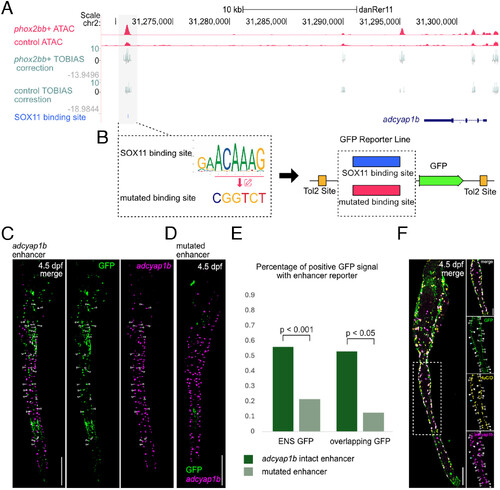
Putative SOX11 binding site in an adcyap1b enhancer. All staining images were after “Max Intensity” z-stack projection. (A) TOBIAS provides Tn5 bias correction of bulk ATAC seq results and SOX11 footprinting. (B) The adcyap1b enhancer with either the intact or mutated SOX11 binding site was cloned into the GFP reporter plasmid. (C) At 4.5 dpf, embryos showed adcyap1b enhancer mediating GFP reporter expression in the gut. Arrows indicate cells coexpressing GFP and adcyap1b. (Scale bar, 100 μm.) (D) At 4.5 dpf, embryos showed a reduction in adcyap1b enhancer-mediated reporter expression after mutation of SOX11 binding sites. (Scale bar, 100 μm.) (E) Enhancers with mutated SOX11 binding site showed a significant reduction in the percentage of positive GFP signal in ENS (ENS GFP, Nadc = 60, Nmut = 51, replicates = 3; overlapping GFP, Nadc = 17, Nmut = 16, replicates = 3). (F) Antibody and HCR staining comparing coexpression of GFP signals, HuC/D and adcyap1b in ENS of Tg(+32adcyap1b:GFP) at 4.5 dpf. The white dash box indicates the zoomed region on the right. Hollow arrows point to GFP+/HuC/D+/adcyap1b+ cells, white arrows point to GFP+/HuC/D+/adcyap1b− cells, and blue arrows point to GFP+/HuC/D−/adcyap1b− cells. (Scale bar, 100 μm, and 50 μm for zoomed region.) |