|
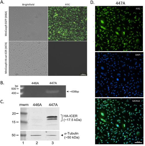
Characterization of established cell lines from fish melanomas. (A) Images of brightfield and fluorescence microscopy of MiniCoopR-EGFP (446A) and MiniCoopR-HA-wt-ICER (447A) cell lines in culture. (B) PCR analysis of 446A and 447A cell lines to test for HA-ICER transgene integration into the fish genome. (C) Anti-HA Western blot analysis of 446A and 447A cell lines to test for transgenic HA-ICER protein expression. 446A was used as a control. (D) Immunocytochemical determination of the subcellular localization of the transgenic HA-ICER protein in 447A cell line. 446A was used as a control, showing no signal (not shown). Commercially available anti-HA antibodies were used in the experiment shown in C and D, and anti-αTubulin antibodies in C as loading control. Scale bar: 50 µm.
|