
Fig. 5
- ID
- ZDB-FIG-250825-65
- Publication
- Ostenrath et al., 2025 - Inhibition mediated by group III metabotropic glutamate receptors regulates habenula activity and defensive behaviors
- Other Figures
- All Figure Page
- Back to All Figure Page
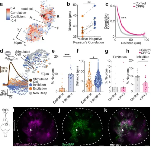
Group III mGluRs play an important role in coordinating spontaneous habenular activity and mediating inhibitory interactions between habenular neurons. a Pearson’s correlations of spontaneous activity between Hb neurons and a seed neuron (green) in Tg(elavl3:GCaMP6s) larval zebrafish. Red means high and blue low correlations. Scale bar represents 20 m. L left, R right, A anterior, P posterior. b Average distance between Hb neuron pairs that are significantly positively or negatively correlated. Positive correlated neurons are significantly closer than negative ones. (n = 7 fish, **p = 0.0035, one sided Wilcoxon signed-rank test). c Pairwise Pearson’s spontaneous activity correlation of habenula neurons as a function of distance ( m) in control (grey) versus 5 mM CPPG-injected fish (pink). CPPG-injected animals exhibit stronger correlations over longer distances. Shadow represents +/-SEM. (Control: n = 14 hemispheres from 7 fish, CPPG-injected: n = 14 Hb hemispheres from 7 fish, ANOVA-n displayed significance over distances ***p = 1.8 × 10⁻¹¹ and over treatment groups ***p = 0.0004). d Calcium imaging of Tg(eval3:GCaMP6-nuclear) juvenile zebrafish brain explant while simultaneously stimulating a single cell via whole-cell recording, brown: stimulated neuron, orange: excited, blue: inhibited, grey: non-responding. Example calcium traces are shown on the left ( F/F). e Percentage of habenula neurons excited or inhibited upon single habenular neuron micro-stimulation. Significantly more cells are inhibited than excited (n = 28 stimulated individual neurons, ***p = 0.00007, two-sided Wilcoxon signed-rank test. f Distance ( m) of the responding neuron (excited, orange or inhibited: blue) to the micro-stimulated neuron. Inhibited neurons are significantly more distant to the stimulated neurons than the excited ones (n = 104 excited neurons and n = 558 inhibited neurons after 28 stimulated neurons, *p = 0.0291, two-sided Wilcoxon signed-rank test)(g, h) Percentage of habenula neurons increasing (Excitation, g) or decreasing (Inhibition, h) their fluorescence upon single cell stimulation during control conditions or during bath application of 300 M CPPG. Significantly less cells are inhibited when 300 M CPPG is applied, but no difference for the fraction of excited neurons. (ACSF n = 28 stimulated neurons; CPPG n = 19 stimulated neurons, Excitation p is n.s; Inhibition **p = 0.007 two-sided Wilcoxon rank sum test). Note that the control data is same as in (e). i–k Confocal microscopy images of tissue-cleared Tg(narp:GAL4VP16;UAS:Synaptophysin–GFP-T2A-tdTomato-CAAX) juvenile zebrafish (n = 5 fish). Colors represent tdTomatoCAAX (magenta) and Synaptophysin–GFP (Syp-GFP, green). Dorsal habenula neurons expressing tdTomatoCAAX (i), Synaptophysin–GFP (j) and merged (k). White line delineate the habenula. Scale bar represents 10 m. White arrow points at Synaptophysin–GFP on the dendritic processes of narp labelled dorsal habenula neurons. A anterior, P posterior. See also Supplementary Fig. 8. Error bars represent mean +/- SEM. Scattered dots represent individual fish (b) or neurons (e–h). Source data are provided as a Source Data file. |