
Fig. 5
- ID
- ZDB-FIG-250822-60
- Publication
- Cakan-Akdogan et al., 2025 - Novel anti-VEGF scFv antibodies with superior in vitro and in vivo activities
- Other Figures
- All Figure Page
- Back to All Figure Page
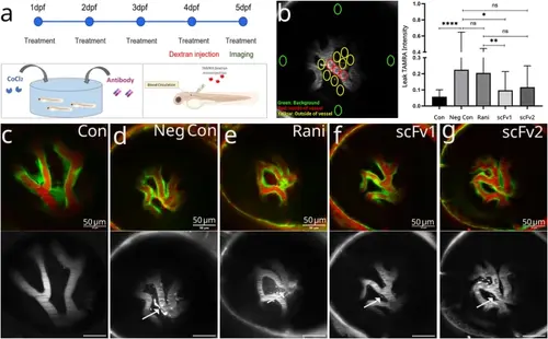
Inhibition of retinal leakage. (A) Graphic representation of the experimental design. Zebrafish embryos were kept in embryo medium containing CoCl2 and antibodies to be tested from 1–5 dpf. TAMRA dextran was injected to the vasculature at 4 dpf larvae and retinal vessels were imaged at 5 dpf. (B) Dye intensity was measured in every retina, in different comparable positions. The number of ROIs and locations are indicated on the image with circles. Larval background signal (green circles), signal in the vein (red circles) and signal around the vessels (yellow circles). Plot shows normalized average leak intensity in each group (n = 5) larvae per group, 2 independent experiments were conducted, similar results were obtained. Results of one experiment is displayed. Statistical significance was tested by one-way ANOVA followed by Kruskal-Wallis comparison test. Error bars represent ± SD ns: non-significant, * P ≤ 0.05, ** P ≤ 0.01, *** P ≤ 0.001, **** P ≤ 0.0001. C - G) Representative images from each experimental group. Overlay image (first row), TAMRA image (second row) is shown for each condition. (C) Control larva that was maintained in normal embryo medium, (D) negative control larva that was treated with CoCl2 but did not receive any anti-VEGF, (E) ranibizumab treated, (F) scFv1 treated and (G) scFv2 treated larva. |