|
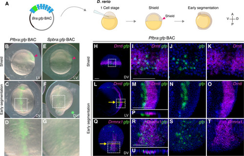
Pfbra:gfp BAC activates GFP expression in the organizer and the hypochord of zebrafish embryos. (A) Circular Pfbra/Spbra:gfp BACs were each introduced into zebrafish zygotes, and GFP signals were observed at the shield and early segmentation stages (A, anterior; P, posterior; V, ventral; D, dorsal). (B to G) GFP signals in zebrafish embryos injected with Pfbra:gfp BAC or Spbra:gfp BAC. GFP signal in the dorsal margin (shield) at the shield stage is indicated by the red arrowhead. The white dashed boxes in (C) and (F) are magnified and shown in (D) and (G), respectively. (H to U) Double fluorescent in situ hybridization of gfp (green) and Drntl [magenta in (H) to (P)] or Drmnx1 (magenta in Q to U) in Pfbra:gfp BAC-injected zebrafish embryos at indicated stages. Nuclei were counterstained with Hoechst 33342 (blue). The areas in the white dashed boxes in (H), (L), and (Q) are enlarged and shown in (I), (M), and (R); single-channel images are shown in (J) and (K), (N) and (O), and (S) and (T), respectively. [(P) and (U)] The XZ sections along the yellow dashed lines are indicated with yellow arrows in (L) and (Q). The orientation of embryos is indicated in each panel: LV, lateral view; DV, dorsal view. All scale bars represent 100 μm. Panels that are in the same scale: (B), (C), (E), and (F); (D) and (G); (H), (L), and (Q); (I) to (K), (M) to (P), and (R) to (U). Created in BioRender. Fan, T. (2025) https://BioRender.com/n8z3595.
|