FIGURE
            Fig. 11
- ID
- ZDB-FIG-250723-11
- Publication
- Milovanovic et al., 2025 - Activation of ankrd1a expression marks newly forming myofibers and regulates muscle cell differentiation in adult zebrafish skeletal muscle repair
- Other Figures
- All Figure Page
- Back to All Figure Page
                
                    
                        Fig. 11


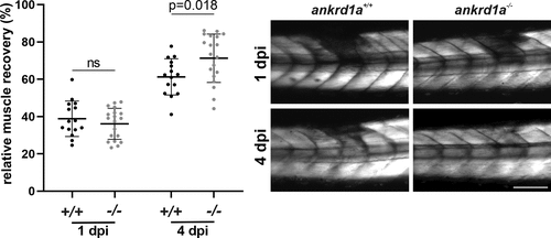
| Relative skeletal muscle recovery in ankrd1a mutant (ankrd1a−/−) and wild-type (wt) (ankrd1a+/+) larvae injured by glass capillary at 72 h postfertilization (hpf). Muscle integrity was analyzed in 15 wt and 19 ankrd1a mutant larvae. Images were taken 1 and 4 days after the injury. Birefringence intensity was measured in LAS X software. Results are expressed as a percentage of signal intensity (gray values/µm2) in injured somites when the signal intensity of adjacent noninjured somites on both sides of the injury was set to 100%. Scale bar, 500 µm. ns, nonsignificant. | 
                
                    
                        Expression Data


                        Expression Detail


                        Antibody Labeling


                        Phenotype Data


                        Phenotype Detail


                        Acknowledgments


This image is the copyrighted work of the attributed author or publisher, and
ZFIN has permission only to display this image to its users.
Additional permissions should be obtained from the applicable author or publisher of the image.
      Full text @ Am. J. Physiol. Cell Physiol.


        