
Fig. 4
- ID
- ZDB-FIG-250721-23
- Publication
- Müller et al., 2025 - Ena/VASP-EVH1 inhibition prevents chemotaxis and metastasis by blocking the EVH1-WAVE2 interaction
- Other Figures
- All Figure Page
- Back to All Figure Page
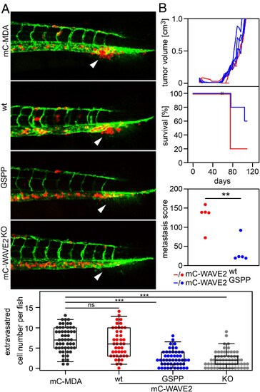
EVH1–WAVE2 interaction enables MDA-MB-231 cell extravasation and metastasis in vivo. (A) Extravasation into the avascular collagen matrix-rich tailfin of zebrafish. Upper panel: micrographs of transgenic zebrafish. Vasculature (green) and mCherry expressing MDA-MB-231 cancer cells (red). White arrows indicate the region of extravasation. Lower panel: statistical evaluation of extravasated cancer cells per fish. Data from three independent experiments, and representative results are shown. Statistical significance (ns: not significant, ***P ≤ 0.001) was evaluated using t test analysis. (B) Xenograft mouse experiment with MDA-MB-231 cells. NOG mice with orthotopically implanted mC-WAVE2wt (red) and mC-WAVE2GSPP (blue) cells. Upper panel: primary tumor volume over time. Middle panel: survival (red and blue lines) with two dropouts per group at day 53 (II). These animals were killed to ensure metastasis development. Lower panel: Metastasis score (red and blue dots). Metastasis score was determined ex vivo after the termination of each animal and represents the sum of all found metastasis in mm2 visible on the surface of the liver and lung measured with a caliper. Statistical significance (**P ≤ 0.01) was evaluated using t test analysis. |