
Fig. 4
- ID
- ZDB-FIG-250714-31
- Publication
- Sepulveda et al., 2025 - Expression of a protein disulfide isomerase A3 variant associated with amyotrophic lateral sclerosis triggers disease features in mice
- Other Figures
- All Figure Page
- Back to All Figure Page
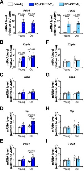
Dysregulation of ER proteostasis in mutant PDIA3Q481K transgenic mice. Lumbar spinal cord tissue of PDIA3Q481K and PDIA3WT transgenic mice and their respective non-transgenic littermates was analyzed for the determination of mRNA levels of ER stress markers at different ages. Young and old correspond to 4 and 18 months of age, respectively. (A) Overexpression of Pdia3 at the mRNA level in the different lines and ages analyzed. (B) Fold change of mRNA levels of the UPR-inducible transcription factor X-box binding protein 1 spliced form (Xbp1s) in the PDIA3Q481K transgenic line. (C) Fold change of mRNA levels of the UPR-inducible transcription factor C/EBP homologous protein (Chop) in the PDIA3Q481K transgenic line. (D) Fold change of mRNA levels of the ER chaperone Binding immunoglobulin protein (Bip) in the PDIA3Q481K transgenic line. (E) Fold change of mRNA levels of the ER oxidoreductase Protein disulfide isomerase a1 (Pdia1) in the PDIA3Q481K transgenic line. (F) to (I), same as (B) to (E) for the PDIA3WT transgenic line. Data are shown as mean ± s.e.m. and statistical analysis was performed using two-tailed Student's t-test. Differences with p < 0.05 were considered statistically significant. If not indicated, comparisons were non-significant. Each symbol in the bar graphs corresponds to an independent biological replicate. |