
Fig. 2
- ID
- ZDB-FIG-250710-67
- Publication
- Zhang et al., 2024 - GPI transamidase complex is required for primordial germ cell migration and development in zebrafish
- Other Figures
- All Figure Page
- Back to All Figure Page
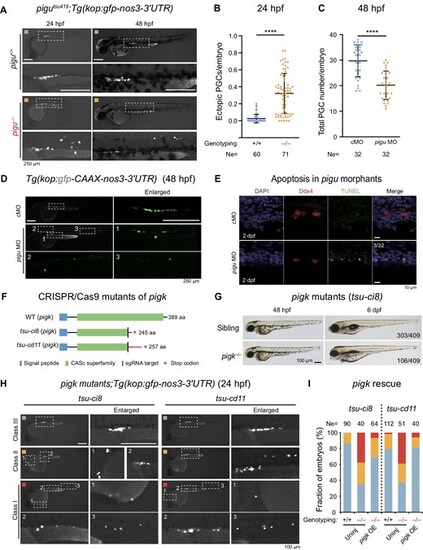
GPIT complex is required for PGC migration. (A) Fluorescence images showing the PGCs in pigu+/+ and pigu–/–embryos by the crossing with Tg(kop:gfp-nos3-3′UTR) transgenic line. The dashed boxed areas were enlarged for better review. Scale bar, 250 μm. (B) Fraction of ectopic PGCs to total PGCs per embryo in embryos in A at 24 hpf. Ne, number of embryos. ****P < 0.0001. (C) Scatter plot showing the total numbers of PGCs per embryo in cMO- and pigu MO-injected embryos at 48 hpf. (D) Fluorescence images showing kop:gfp signals (PGCs) in cMO- and pigu MO-injected embryos at 48 hpf. Scale bar, 250 μm. (E) Fluorescence images showing cMO- and pigu MO-injected embryos at 12-somite and 2 dpf stages with the TUNEL assay (green) and Ddx4 immunostaining (red), respectively. DAPI, DNA, blue. Scale bar, 10 μm. (F) Schematic showing protein structures of WT and two pigk mutant lines, tsu-ci8 and tsu-cd11. (G) Morphology of pigk mutant embryos at 48 hpf and 6 dpf. The ratios of embryos are shown in the picture. Scale bar, 100 μm. (H) Fluorescence images showing the PGCs in pigktsu-ci8 and pigktsu-cd11 embryos by the crossing with Tg(kop:gfp-nos3-3′UTR) transgenic line in class I (red), class II (orange), and class III (blue), which indicate severe and mild defective and normal PGC migration, respectively. The dashed boxed areas were enlarged for better review. Scale bar, 100 μm. (I) The ratios of embryos in class I (red), class II (orange), and class III (blue) in uninjected WT, pigk mutant, and 200 pg WT pigk mRNA-injected pigk mutant embryos. |