|
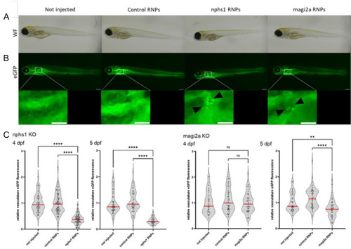
Evaluation of proteinuria in nphs1 and magi2a KO embryos. A shows the general morphology of larvae which were either uninjected, injected with control RNPs, or with nphs1 or magi2a RNPs. The fluorescence microcraphs in B show the filtered and reabsorbed gc-eGFP fusion protein in proximal tubules, but not in control- or uninjected embryos. As shown in C, in the beforehand established high-content proteinuria screening approach, nphs1 KO embryos were proteinuric at day 4 and 5 as demonstrated by decreased intravascular gc-eGFP fluorescence, while magi2a KO embryos were significantly proteinuric later at day 5. Scale bars indicate 200 µm in the overview and 100 µm in the zoom inserts
|