IMAGE

Fig. 9

ID
ZDB-IMAGE-250627-111
Source
Figures for Siegerist et al., 2025
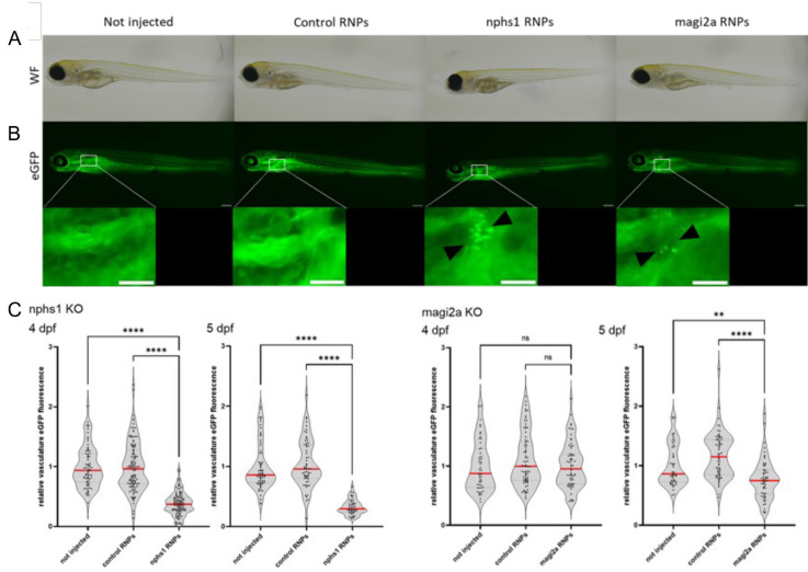
Image
Figure Caption

Fig. 9

Evaluation of proteinuria in nphs1 and magi2a KO embryos. A shows the general morphology of larvae which were either uninjected, injected with control RNPs, or with nphs1 or magi2a RNPs. The fluorescence microcraphs in B show the filtered and reabsorbed gc-eGFP fusion protein in proximal tubules, but not in control- or uninjected embryos. As shown in C, in the beforehand established high-content proteinuria screening approach, nphs1 KO embryos were proteinuric at day 4 and 5 as demonstrated by decreased intravascular gc-eGFP fluorescence, while magi2a KO embryos were significantly proteinuric later at day 5. Scale bars indicate 200 µm in the overview and 100 µm in the zoom inserts

Acknowledgments
This image is the copyrighted work of the attributed author or publisher, and ZFIN has permission only to display this image to its users. Additional permissions should be obtained from the applicable author or publisher of the image. Full text @ J Transl Med