|
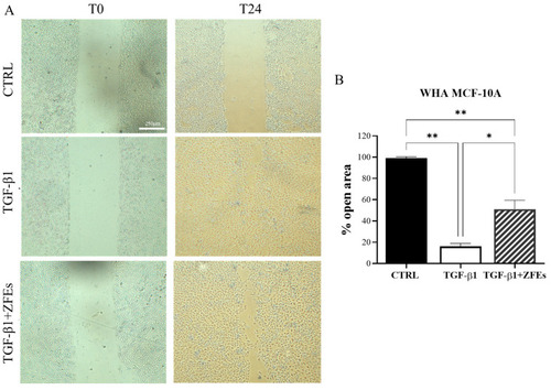
Wound-healing assay in MCF-10A cells. (A) Representative phase-contrast images of the wound-healing assay taken 24 h post-scratch. Images were obtained via optical microscopy. (B) Quantitative analysis of wound closure after scratch. Normal breast cells, upon TGF-β1 treatment, exhibited migratory properties, almost completely filling the scratch area. However, ZFEs treatment reduced the migratory rate, with more than 50% of the area remaining open. Values are expressed as the mean percentage of residual open area compared to the respective cell-free gap at T0. Data are presented as mean ± SD. Statistical analysis was conducted using one-way ANOVA followed by Tukey’s multiple comparison test. * p < 0.05; ** p < 0.01. Scale bars: 250 μm.
|