|
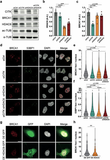
HDAC6 targeting recovers BRCA1 levels and DNA damage in CITK depleted cells. a Western blot analysis of total lysate from ONS-76 cells, 48 hafter transfection of the indicated siRNAs. The levels of CITK, HDAC6, acetylated tubulin (ac-TUB) were analyzed, and the internal loading control was α-tubulin (α-TUB). b Quantification of the relative density of HDAC6 in each treatment condition. c Quantification of the relative density of BRCA1 in each treatment condition. d Representative images of ONS-76 cells, analyzed 48 h after transfection of the indicated siRNAs. Cells were immunostained for BRCA1 and 53BP1 and counterstained with DAPI. Scale bars: 10 μm. Quantification of BRCA1 (e) or 53BP1 (f) foci per nucleus in experiments performed as in (d). g Representative images of ONS-76 cells after transfection with GFP or HDAC6-GFP expression plasmids. Cells were immunostained for BRCA1 and counterstained with DAPI. Scale bars: 10 μm. h Quantification of BRCA1 foci per nucleus in samples treated as in (e) condition. Each dot indicates an independent biological replicate. All immunofluorescence quantifications were based on at least four independent biological replicates; >300 cells were analyzed per condition in each replicate. Error bars, SEM. **P < 0.01, ***P < 0.001, ****P < 0.0001; one-way ANOVA test followed by Bonferroni’s correction (b, c, e, f), unpaired Student’s t-test (h). A.U. arbitrary unit.
|