
Fig. 4
- ID
- ZDB-FIG-250421-59
- Publication
- Zhu et al., 2024 - Receptor binding and tortuosity explain morphogen local-to-global diffusion coefficient transition
- Other Figures
- All Figure Page
- Back to All Figure Page
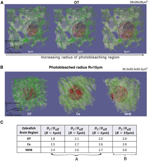
FRAP simulations on different photobleaching radius sizes. (A) Simulations of FRAP with photobleaching regions of spheres with different radius sizes, μm (left to right). μm3 tissue segments were used for FRAP simulations. (B) Full 3D image of FRAP on OT, Ce, and MHB regions (left to right), where the photobleaching region was μm. μm3 tissue segments were used for these sizes of FRAP simulation. This shows that the tissue structure varies across zebrafish brain regions. (C) Values of from FRAP experiments for the various radius sizes, showing that we obtain a factor of around 2–3 decrease of effective diffusion coefficient for various zebrafish brain regions. The values show that FRAP results may slightly vary depending on the tissue architecture surrounding the photobleached region. |