FIGURE

Figure 8

ID
ZDB-FIG-250417-122
Publication
Derrick et al., 2024 - Zebrafish arterial valve development occurs through direct differentiation of second heart field progenitors
Other Figures
All Figure Page
Back to All Figure Page
Figure 8

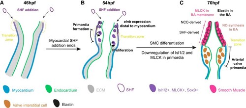
The zebrafish arterial valve forms by direct differentiation of SHF progenitors. Model of zebrafish arterial valve formation. (A) At 46 hpf, addition to the OFT from the SHF occurs at the transition zone, where cells co-express Isl1/2 and mature cardiomyocyte markers, before down-regulating Isl1/2. (B) At 54 hpf, SHF addition no longer adds myocardium to the OFT, but instead smooth muscle which expresses elnb. This switch is the site of formation of the primordia (the transition zone), forming a bulge between the myocardium and endocardium. Cells within the early primordia proliferate and share similar expression patterns to the distal smooth muscle, but do not express elnb. (C) By 70 hpf, the smooth muscle cells of the bulbus arteriosus are now more molecularly distinct from the cells of the arterial valve primordia. The smooth muscle expresses elnb, MLCK, Tagln, and Sox9, is producing NO, and is surrounded by elastin fibres. The interstitial cells of the arterial valve primordia have down-regulated Isl1/2 and MLCK but maintain Sox9. The primordia are devoid of cells expressing elnb or any elastin fibres.

Expression Data

Expression Detail
Antibody Labeling
Phenotype Data

Phenotype Detail
Acknowledgments
This image is the copyrighted work of the attributed author or publisher, and ZFIN has permission only to display this image to its users. Additional permissions should be obtained from the applicable author or publisher of the image. Full text @ Cardiovasc. Res.