FIGURE SUMMARY
Title

Zebrafish arterial valve development occurs through direct differentiation of second heart field progenitors

Authors
Derrick, C.J., Eley, L., Alqahtani, A., Henderson, D.J., Chaudhry, B.
Source
Full text @ Cardiovasc. Res.

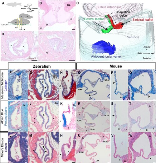
The zebrafish arterial valve is anatomically similar to other vertebrate arterial valves. (A) Overview of the orientation of the heart in adult zebrafish, with planes of imaging for C-N. (B) Haematoxylin and eosin–stained long-axis sections of excised adult zebrafish heart (female, 9 months old) (n = 4). (C) 3D reconstruction of the arterial pole, viewed ventrally, reconstructed from (B). The arterial valve leaflets (green, red) are hinged in the ventricular myocardium and span the myocardial (grey)-arterial (pink) boundary and are not in continuity with the atrioventricular valve (blue). The arterial valve leaflets are aligned along the anterior-posterior axis of the zebrafish and are defined by their position within the adult: dextral (green) and sinistral (red) with their coaptation region spanning the myocardial-arterial boundary. (D and E) Haematoxylin and eosin–stained short-axis sections of excised adult zebrafish heart (male, 9 months old) at the level of distal (D) and proximal myocardium (E) showing two leaflets of the arterial valve, with sinuses (asterisks) between the leaflets and the wall (n = 4). (F) Masson’s trichrome-stained short-axis section of excised adult zebrafish heart (female, 14 months) at the commissures of arterial leaflets in the bulbus arteriosus. Collagen (blue) is present in the bulbus arteriosus, enriched in the commissures and largely absent from the leaflets. (G) Same heart as (F) at the proximal myocardium. Collagen is present in the lumen facing portion of the leaflets (arrowheads), the wall of the sinus (asterisks) but is excluded from the leaflet interstitium. (H) Masson’s trichrome-stained long-axis section of excised adult zebrafish heart (female, 12 months old) showing the sinistral leaflet. Collagen is present on the luminal surface (arrowhead) of the leaflet, the root, and the bulbus arteriosus. (I) Alcian blue–stained sections, same heart shown in (F) and (G). Proteoglycans (blue) are abundant in the bulbus arteriosus and leaflets but absent from the commissures (asterisks). (J) Same heart as (I) at the proximal myocardium. Proteoglycans are present throughout the interstitium of the leaflets, with very weak signal in the sinus wall (asterisk). (K) Alcian blue–stained section, same heart shown in (H), showing the bulbus arteriosus and leaflets are rich in proteoglycans. (L) Miller’s elastin-stained sections, same heart shown in (F), (G), (I), and (J). Elastin (purple) is abundant in the bulbus arteriosus, absent from the tips of the leaflets and commissures (asterisks). (M) Same heart as (L) at the proximal myocardium. Elastin is present throughout the leaflet, but is enriched on the luminal surface of the leaflets and the sinus wall (arrowheads). (N) Miller’s elastin-stained section, same heart shown in (H) and (K). Elastin is present in the bulbus arteriosus, enriched in the lumen facing aspect of the arterial leaflets, and present in the root of the valve (for F, G, I, J, L, and M: n = 5; for H, K, and N: n = 4). (O) Masson’s trichrome-stained short-axis section of postnatal day (P) 90 mouse aortic valve. The aorta and commissures (asterisks) are rich in collagen, whilst there is enrichment on the sinus facing aspect of the leaflets (arrowhead). (P and Q) Masson’s trichrome-stained long-axis section P90 mouse aortic valve, showing left (P) and non-coronary leaflets (Q). Collagen is present in the aortic root, hinge, and wall. In the leaflets, collagen is mainly localized to the arterial aspect and absent from the luminal surface (P and Q, arrowheads). (R) Alcian blue–stained section of same heart shown in (O). Proteoglycans are present in the leaflets and commissures (asterisks) but absent from the aorta. (S and T) Alcian blue–stained section, same heart shown in (P) and (Q). Sulfated proteoglycans are present in the aortic root and hinge, but largely absent for the wall of the aorta. (U) Miller’s elastin-stained section, same heart shown in (O) and (R). Elastin fibres are present in the aorta and absent from the commissures (asterisks) and valve leaflets. (V and W) Miller’s elastin-stained section, same heart shown in (P), (Q), (S), and (T). Mature elastin fibres are present in the wall of the aorta, with diffuse staining in the hinge. There are no clear fibres of elastin in the leaflets. (B), (H), (K), (N), (P), (Q), (S), (T), (V), and (W): anterior: up, left: right. (D)–(G),( I), (J), (L), and (M): dorsal: up, left: right. V, ventricle; BA, bulbus arteriosus; A, atrium; SV, sinus venosus; LV, left ventricle; Ao, aorta; H, hinge; L, lumen; RΔNC, right/non-coronary commissure; NCΔL, non-coronary/left commissure; LΔR, left/right commissure. Scale bars: (B) and (D)–(N): 20 μm; (O)–(W): 50 μm.

Development of the zebrafish arterial valve follows conserved events. (A) Schematic of 30 hpf OFT. Two cell layers, the wall (black) and endocardium (green), are separated by ECM (grey). (A′) Representative haematoxylin and eosin–stained midline section through the long axis of the OFT at 30 hpf; arrow denotes direction of blood flow (n = 6). (BC′) Between 38 and 46 hpf, the two cell layers remain distinct and separated by ECM (38 hpf, n = 7; 46 hpf, n = 10). (D and D′) At 54 hpf, a multi-layering of cells is present at the distal-most point of the OFT where no ECM is visible (D′, arrowhead), with cells of unknown fate in yellow (n = 8). (E and E′) ECM in the ventricle and OFT is largely absent at 62 hpf (n = 11). (FG′). Between 70 and 78 hpf, any remaining ECM is lost and the OFT lengthens and tapers where it connects to the ventral aorta (70 hpf, n = 5; 78 hpf, n = 10). (H and H′) Sinuses are visible by 86 hpf, defining two leaflets in the OFT (arrowheads) (n = 5/8). (I–L) Live fluorescent imaging of the OFT between 62 and 86 hpf of Tg(kdrl:GFP) embryos, marking the endocardium. Two primordia are visible at 62 hpf (I) (n = 13) and 70 hpf (J) (n = 14). The beginning of sinus formation is detectable at 78 hpf (K) (n = 11/15), with tips of leaflets visible (arrowheads) and is mostly complete by 86 hpf (L) (n = 17). V, ventricle; BA, bulbus arteriosus. Scale bars: (A)–(H): 20 μm; (I)–(L): 5 μm.

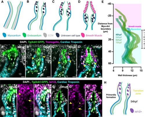
Arterial valve primordia form at the transition zone. (A–D) Schematics of OFT at 46 hpf (A), 54 hpf (B), 62 hpf (C), and 70 hpf (D). (A′–D′) Representative midline sections of Tg(kdrl:GFP) (endocardium, green) embryos stained for Tagln (smooth muscle, magenta), cardiac troponin (myocardium, cyan), and DAPI (white) between 46 and 70 hpf. At 46 hpf (A and A′), the entire OFT is myocardial (n = 12); at 54 hpf (B and B′), there is a region distally that is not myocardial and does not express Tagln (asterisk); some cells are identifiable between the endocardium and myocardium (arrowheads) (n = 15). By 62 hpf (C and C′), the region distal to the myocardium begins to express Tagln (n = 9), which is more robust; at 70 hpf (D and D′) (n = 11), this is the bulbus arteriosus. Cells present between the endocardium and myocardium do not express any markers (C′ and D′ arrowheads). (E) Quantification of wall thickness, averaged across left and right, with distance along OFT measured relative to myocardial-arterial boundary (46 hpf, n = 24; 54 hpf, n = 30; 62 hpf, n = 18; 70 hpf, n = 22). The primordia of the arterial valve form at the distal-most point of the ventricular myocardium. (FG″) Representative midline resin section of Tg(kdrl:GFP) (green), stained for cardiac troponin (cyan), Isl1/2+ cells (SHF, magenta), and DAPI (white) at 46 hpf (FF″, n = 14) and 54 hpf (GG″, n = 14). (FF″) At 46 hpf, both cardiomyocytes (arrowhead) and endocardium (arrow) at the distal end of the OFT are Isl1/2+ (n = 14). (GG″) At 54 hpf, cardiomyocytes (arrowhead) and endocardium (arrow) of the distal OFT are Isl1/2+, whilst cells of the forming primordia are Isl1/2 (asterisk), but do not express myocardial or endocardial markers. The forming bulbus arteriosus is also SHF derived. (H) Summary of expression data, confirming the presence of the transition zone at the arterial pole (yellow region) as the site of primordia formation. E, mean ± SEM; V, ventricle; BA, bulbus arteriosus. Scale bars: 10 μm.

Direct differentiation of SHF progenitors establishes the zebrafish arterial valve. (AB″) Representative midline section of immunohistochemistry on Tg(kdrl:GFP) (green) embryo at 54 hpf (AA″) and 70 hpf (BB″) for cardiac troponin (white, membrane), DAPI (white, nuclear) (A and B), Islet1/2 (Isl1/2, magenta) (A′ and B′), and Sox9 (cyan) (A″ and B″). (AA″) At 54 hpf, cells of the distal OFT, including within the arterial valve primordia (yellow) co-express Isl1/2 and Sox9 (n = 19). (BB″) By 70 hpf, cells within the arterial valve primordia (yellow) have down-regulated Isl1/2 (B′) but maintain expression of Sox9 (B″) defining them as VICs (n = 12). (CD″) Representative midline sections of lineage tracing of endothelial cells in the OFT of Tg(kdrl:GFP); Tg(kdrl:Cre); Tg(-3.5ubb:loxP-EGFP-loxP-mCherry) embryos (magenta) at 70 hpf (CC″) and 118 hpf (DD″). (CC″) Cells of the arterial valve primordia at 70 hpf (yellow), identified between distal-most myocardium (cyan) and endocardium (green), are not of endocardial origin (n = 16). (DD″) At 118 hpf following primordia remodelling, there is no recombination observed in the arterial valve leaflets (yellow, arrowheads denote tips of leaflets) (n = 14). (EF″) Representative midline sections of lineage tracing of neural crest cells in the OFT of Tg(kdrl:GFP); Tg(sox10:iCre, cryaa:DsRed2); Tg(-3.5ubb:loxP-EGFP-loxP-mCherry) embryos (magenta) at 70 hpf (EE″) and 118 hpf (FF″). (EE″) Cells of the arterial valve primordia at 70 hpf (yellow), identified between distal-most myocardium (cyan) and endocardium (green), are not of neural crest origin (n = 12/14). (FF″) At 118 hpf following primordia remodelling, there is no recombination observed in the arterial valve leaflets (yellow) (n = 8/12, arrowheads denote tips of leaflets). At both stages, recombination is present in the distal tip of the bulbus arteriosus (E′ and F′, asterisk). (G) Summary of expression data from (A–F). V, ventricle; BA, bulbus arteriosus. Scale bars: 10 μm.

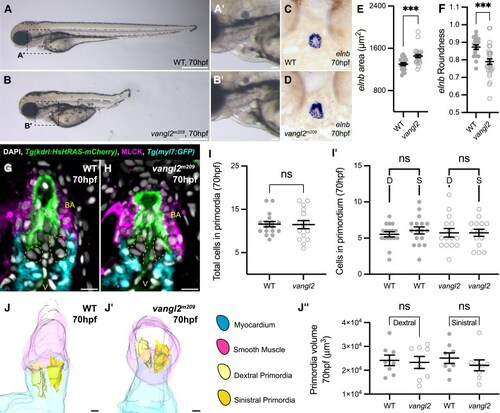
Arterial valve primordia cells are distinct from smooth muscle cells. (A–D) Representative mRNA in situ hybridization analysis of elastin b (elnb) expression during arterial valve primordia formation. elnb is not expressed at 46 hpf (A) (n = 17) and initiates at 54 hpf (B, arrowhead) (n = 17), and the domain expands between 62 and 70 hpf (C–D) (62 hpf, n = 17; 70 hpf, n = 17). (E–H) Midline resin sections of embryos stained for elastin fibres by Miller’s elastin between 46 and 70 hpf. No elastin fibres are present at 46–54 hpf (E and F) (46 hpf, n = 13; 54 hpf, n = 12). Fibres are present at 62 hpf (G) (n = 11), and by 70 hpf (H, n = 12), the majority of the bulbus arteriosus has elastin fibres, but these are not present in the primordia (arrowheads in FH). (IJ″) Representative midline resin section of immunohistochemistry on Tg(kdrl:HsHRAS-mCherry) (magenta) embryos incubated with the NO sensor DAF-FM (green) and stained with cardiac troponin (cyan) and DAPI (white) at 54 hpf (II″) and 70 hpf (JJ″). At 54 hpf, there is weak, sparse DAF-FM staining of the putative SHF-derived smooth muscle (n = 13/18), and there is no staining in the arterial primordia (yellow region). At 70 hpf (JJ″), DAF-FM staining is present throughout the majority of the bulbus arteriosus, with some cells negative at the distal-most point. There is no staining of the arterial valve primordia (yellow region) (n = 13). (KL″) Representative midline resin section of immunohistochemistry on embryos incubated with the NO sensor DAF-FM (green) and stained with cardiac troponin (cyan), MLCK (magenta), and DAPI (white) at 54 hpf (KK″) and 70 hpf (LL″). As in (I′), at 54 hpf, there is weak, sparse DAF-FM staining of the putative SHF-derived smooth muscle and no staining in the arterial primordia (yellow region) (n = 11/14). In all embryos, MLCK signal is present in the valve primordia and undifferentiated SHF. At 70 hpf (LL″), MLCK signal is membrane restricted, absent from the arterial primordia and marks the entirety of the bulbus arteriosus (arrowhead), overlapping with DAF-FM staining (n = 14). V, ventricle; BA, bulbus arteriosus. Scale bars: 10 μm.

tbx1 mutants have a dysmorphic OFT and hypocellular arterial valve primordia. (AB′) Representative brightfield image of WT sibling (A, n = 6) and tbx1tm208 homozygous mutant (B, n = 12) at 70 hpf, dashed boxes in (A) and (B) highlights heart shown in (A′) and (B′). In tbx1tm208 mutants (B′), the pharyngeal arches are absent (asterisk), and the angle of the OFT angle is steeper than in WT (white). (CD″) Representative images of mRNA in situ hybridization for elnb in WT sibling (C) and tbx1tm208 homozygous mutants (DD″) at 70 hpf. tbx1 mutants display variability in size of elnb domain. (E and F) Representative midline sections of immunohistochemistry on WT sibling (E) and tbx1tm208 homozygous mutants (F) carrying Tg(fli1a:AC-TagRFP) to mark the endocardium (green), cardiac troponin (cyan), and MLCK (magenta). The tbx1tm208 OFT is smaller and dysmorphic with diffuse MLCK expression. (G and G′) Quantification of number of cells in arterial valve primordia (yellow, E and F) at 70 hpf in WT sibling and tbx1tm208 homozygous mutants at 70 hpf. Loss of tbx1 results in a significant reduction in number of VICs and impacts the dextral (D) and sinistral (S) primordia equally (WT sibling, n = 13; tbx1tm208, n = 12). (G) and (G′): Mean ± S.E.M, Welch’s unpaired t-tests. ****, P < 0.0001; ns, not significant. V, ventricle; BA, bulbus arteriosus; D, dextral; S, sinistral. Scale bars: (A) and (B): 500 μm; (A′) and (B′): 50 μm; (C)–(D′): 20 μm; (E) and (F): 10 μm.

vangl2 mutants have misshapen OFT. (AB′) Representative brightfield image of WT sibling (A, n = 7) and vangl2m209 homozygous mutant (B, n = 15) at 70 hpf, dashed boxes in (A) and (B) highlights heart shown in (A′) and (B′). The overt morphology of the vangl2m209 OFT appears normal (white). (C and D) Representative images of mRNA in situ hybridization for elnb in sibling (C) and vangl2m209 homozygous mutant (D) at 70 hpf. (E) Quantification of area of elnb domain at 70 hpf. Loss of vangl2 results in a larger elnb domain. (F) Quantification of roundness of elnb domain at 70 hpf. Loss of vangl2 leads to a less round elnb domain (WT sibling, n = 19; vangl2m209, n = 28). (G and H) Representative midline sections of immunohistochemistry on WT sibling (G) and vangl2m209 homozygous mutants (H) carrying Tg(kdrl:HsHRAS-mCherry) to mark the endocardium (green) and Tg(myl7:GFP) to mark the myocardium (cyan) and MLCK (magenta). The vangl2 OFT appears squat, with the myocardial collar less pronounced. (I and I′) Quantification of number of cells in arterial valve primordia (yellow) at 70 hpf in WT sibling and vangl2m209 homozygous mutants at 70 hpf. Loss of vangl2 does not impact number of VICs ((WT sibling, n = 17; vangl2m209, n = 15), D, dextral primordia; S, sinistral primordia). (JJ″) Quantification of arterial valve primordia volume by 3D reconstruction of WT sibling (J) and vangl2m209 (J′) homozygous mutant OFT from immunohistochemistry. Loss of vangl2 does not impact primordia volume (J″). (E), (F), (I), and (I′): Mean ± SEM, Welch’s unpaired t-tests. (J″): Mean ± SEM, Brown–Forsyth and Welch’s ANOVA. ***, P < 0.001; ns, not significant. V, ventricle; BA, bulbus arteriosus; D, dextral; S, sinistral. Scale bars: (A) and (B): 500 μm; (A′) and (B′): 50 μm; (C), (D), (J), and (J′): 20 μm; (G) and H): 10 μm.

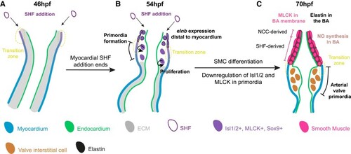
The zebrafish arterial valve forms by direct differentiation of SHF progenitors. Model of zebrafish arterial valve formation. (A) At 46 hpf, addition to the OFT from the SHF occurs at the transition zone, where cells co-express Isl1/2 and mature cardiomyocyte markers, before down-regulating Isl1/2. (B) At 54 hpf, SHF addition no longer adds myocardium to the OFT, but instead smooth muscle which expresses elnb. This switch is the site of formation of the primordia (the transition zone), forming a bulge between the myocardium and endocardium. Cells within the early primordia proliferate and share similar expression patterns to the distal smooth muscle, but do not express elnb. (C) By 70 hpf, the smooth muscle cells of the bulbus arteriosus are now more molecularly distinct from the cells of the arterial valve primordia. The smooth muscle expresses elnb, MLCK, Tagln, and Sox9, is producing NO, and is surrounded by elastin fibres. The interstitial cells of the arterial valve primordia have down-regulated Isl1/2 and MLCK but maintain Sox9. The primordia are devoid of cells expressing elnb or any elastin fibres.

Acknowledgments
This image is the copyrighted work of the attributed author or publisher, and ZFIN has permission only to display this image to its users. Additional permissions should be obtained from the applicable author or publisher of the image. Full text @ Cardiovasc. Res.