
Fig. 6
- ID
- ZDB-FIG-250416-92
- Publication
- Jia et al., 2025 - Activation of prep expression by Tet2 promotes the proliferation of bipotential progenitor cells during liver regeneration
- Other Figures
- All Figure Page
- Back to All Figure Page
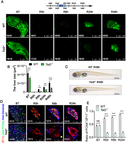
Tet2 mutant blocks liver regeneration. (A) Confocal microscope projection images showing the size of the regenerating livers of WT siblings and Tet2−/− at BT, R0h, R8h, R24h and R48h. The observed green fluorescence is derived from the Denra2 fluorescent protein, specifically labeling the liver of zebrafish. (B) Area of fluorescence statistics of Dendra2 for A. (C) Brightfield and epifluorescence merged images showing the body phenotypes of WT siblings and Tet2−/− at R48h. (D) Single-section confocal microscope images showing anti-PCNA and anti-2F11 antibody staining of the regenerated livers of WT siblings and Tet2−/− at BT, R0h, R8h, R24h and R48h. (E) Ratio of PCNA+ cells to 2F11+ cells in regenerated livers from C. ns, not significant; *P<0.05, **P<0.01, ***P<0.001 (two-tailed post hoc test). Data are mean±s.e.m. Scale bars: 100 µm (A); 500 µm (C); 75 µm (D). |