
Fig. 5
- ID
- ZDB-FIG-250416-91
- Publication
- Jia et al., 2025 - Activation of prep expression by Tet2 promotes the proliferation of bipotential progenitor cells during liver regeneration
- Other Figures
- All Figure Page
- Back to All Figure Page
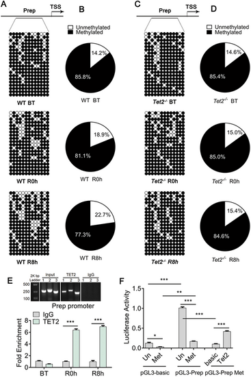
Tet2 activates prep expression. (A) Bisulfite sequencing analysis of DNA methylation at 13 independent CpG islands in the prep promoter locus in DNA isolated from WT regenerated livers at BT, R0h and R8h. Top panel: scheme illustrating the prep promoter region (−890 bp to −1390 bp) containing the 13 independent CpG island sites. TSS, transcription start site. Bottom panel: circle diagram showing the results of DNA methylation at 13 independent CpG islands in the prep promoter region. (B) Sector plots indicating the DNA methylation statistics of 13 independent CpG sites in the prep promoter region of WT regenerated livers at BT, R0h and R8h. The DNA demethylation of the prep promoter region was enhanced during liver regeneration. (C) Bisulfite sequencing analysis of DNA methylation at 13 independent CpG islands in the prep promoter locus in DNA isolated from Tet2 homozygous mutant (Tet2−/−) regenerated livers at BT, R0h and R8h. (D) Sector plots indicating the DNA methylation statistics of 13 independent CpG sites in the prep promoter of Tet2−/− regenerated livers at BT, R0h and R8h. The DNA demethylation of the prep promoter region was inhibited in Tet2−/− [open rings represent ‘unmethylated’ and filled rings represent ‘methylated’ in A and C, and the percentage statistics for unmethylated (white) and methylated (black) in B and D are from A and C]. (E) Top panel: anti-TET2 and anti-IgG antibody chromatin immunoprecipitation (ChIP) DNA electrophoresis showing a Prep promoter region DNA fragment size of approximately 250 bp. Bottom panel: ChIP-qPCR assays of anti-TET2 and anti-IgG antibody in DNA isolated from WT regenerated livers at BT, R0h and R8h. Results showed that the prep promoter was significantly increased in the anti-TET2 group compared with the anti-IgG control group. (F) Luciferase activity assay showed that the transcriptional activity of the unmethylated pGL3-Prep promoter was increased compared with the methylated pGL3-Prep promoter; adenovirus-transfected 293T cell overexpression of the TET2 protein enhanced transcriptional activity of the methylated pGL3-Prep promoter. *P<0.05, **P<0.01, ***P<0.001 (two-tailed post hoc test). Data are mean±s.e.m. |