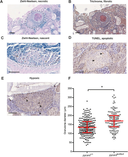
pycardtpu4/tpu4 fish display an increase in granuloma size. Six wild-type (WT) and four pycardtpu4/tpu4 fish were infected with a low dose (mean, 64 CFU; range, 53-76 CFU) of M. marinum. At 8 wpi, the fish were processed for histological analysis of granulomas with Ziehl–Neelsen stain, Mallory's trichrome stain, hypoxia staining and TUNEL assay. (A) A necrotic and fibrotic granuloma stained with Ziehl–Neelsen stain. Dashed line indicates the area that was considered to belong to the capsule of the granuloma. (B) A necrotic and fibrotic granuloma stained with Mallory's trichrome stain. (C) A nascent granuloma, Ziehl–Neelsen staining. (D) A granuloma containing apoptotic cells, TUNEL assay. (E) A hypoxic granuloma. Scale bars: 50 µm. In B-E, dashed line indicates the borders of the granuloma; arrowheads indicate the positively stained cells. (F) The numbers and characteristics of each granuloma [n=244 (pycard+/+), n=128 (pycardtpu4/tpu4)] were recorded for each fish. Using a linear mixed model, granulomas in pycardtpu4/tpu4 were determined to be larger than those in WT siblings (*P=0.0217) using R-package lme4, with fish as a random and genotype as a fixed factor. The line indicates the median and the interquartile range. Only male fish were used as female fish often present an increased number of small granulomas in the gonads, which complicates analyses. See also Fig. S5 for sizes of individual fish granulomas and Fig. S6 for characterization.
|
