
Fig. 3
- ID
- ZDB-FIG-250407-12
- Publication
- Xu et al., 2025 - The synergistic effect of c-Myb hyperactivation and Pu.1 deficiency induces Pelger-Huët anomaly and promotes sAML
- Other Figures
- All Figure Page
- Back to All Figure Page
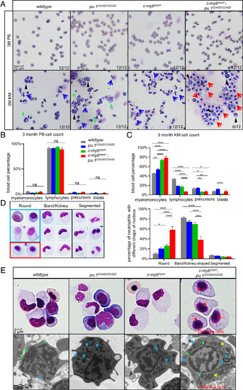
The myeloid cell count increased in 3-mo-old c-mybhyper;pu.1G242D/G242D zebrafishKM with neutrophils showing Pelger–Huët anomaly–like changes. (A–C) PB cells and KM blood cells (A) in 3-mo-old adult fish stained with May-Grunwald/Giemsa [400×, (black triangles indicate blasts, green triangles precursors, blue arrows indicate normal band/kidney-shaped neutrophils, and red arrows indicate abnormally rounded neutrophils)]. Blood cell counts of PB (B) and KM (C) were calculated manually based on their morphology. The black asterisks indicate statistical difference (n = 12). (D) Morphology and counting ratio of neutrophils with different nuclear morphologies (400×, the red box shows abnormal round nuclear cells in c-mybhyper;pu.1G242D/G242D zebrafish, the blue box shows normal immature round nuclear cells from the other three genotypes n = 6). (E) High-resolution imaging (upper, 600×) and TEM morphology (Lower) observation showed PHA-like cells in c-mybhyper;pu.1G242D/G242D zebrafish. Blue, yellow, red, and green arrows indicate crystal granules, vacuolar granules, chromatin clumping, and nuclear membranes, respectively. All TEM images are magnification ×12,000 and 80 kV. |