|
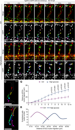
High glucose treatment caused excessive migration of vascular endothelial cell (EC) nuclei in intersegmental vessels (ISVs) of zebrafish embryos. A through N, Still images from in vivo time-lapse imaging analysis of Tg(fli1a:nGFP::kdrl:ras-mCherry) embryos from 63 to 75 hours post-fertilization (hpf). A′ through N′, The nGFP channel of the above panels, respectively. The numbers represent different EC nuclei in unilateral ISVs. O and P, Diagrams of the migration routes of EC nuclei labeled in A′ through N′. Q, Statistics of the distance of EC nuclei migration at different time stages in control (Ctrl) embryos (n=8) and high glucose–treated embryos (n=8). Two-way ANOVA. R, The number of EC nuclei in different migration distances of Ctrl embryos and high glucose–treated embryos. Scale bars, 50 µm.
|