
FIGURE
Fig. 1 - Supplemental 3
- ID
- ZDB-FIG-250218-66
- Publication
- Debaenst et al., 2025 - Crispant analysis in zebrafish as a tool for rapid functional screening of disease-causing genes for bone fragility
- Other Figures
- All Figure Page
- Back to All Figure Page
Fig. 1 - Supplemental 3
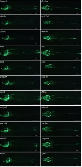
Osteoblast-positive head area at 14 dpf. Visualization of the osteoblast using the osx:Kaede transgenic line. The first four genes are associated with the pathogenesis of osteoporosis, while the last six are linked to osteogenesis imperfecta. The presented image shows a representative image of the specific crispants. Images are taken with the Leica microscope and the osx:Kaede-positive larvae are visualized from a ventral and lateral perspective. Scale bars = 1 mm. |
Expression Data
Expression Detail
Antibody Labeling
Phenotype Data
Phenotype Detail
Acknowledgments
This image is the copyrighted work of the attributed author or publisher, and
ZFIN has permission only to display this image to its users.
Additional permissions should be obtained from the applicable author or publisher of the image.
Full text @ Elife