
Fig. 1
- ID
- ZDB-FIG-250218-63
- Publication
- Debaenst et al., 2025 - Crispant analysis in zebrafish as a tool for rapid functional screening of disease-causing genes for bone fragility
- Other Figures
- All Figure Page
- Back to All Figure Page
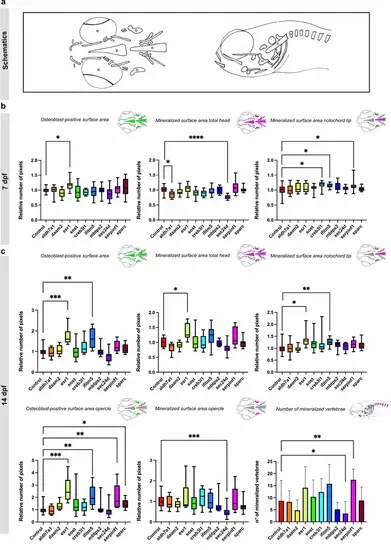
Measurements of osteoblast-positive surface area and mineralized surface area of the head skeleton in 7 and 14 dpf crispants. The first four genes are associated with the pathogenesis of osteoporosis, while the last six are linked to osteogenesis imperfecta. (a) Schematic overview of the ventral and lateral perspective of the head of zebrafish larvae at 7 and 14 dpf. The notochord tip (n), the opercle (o) the mineralized vertebrae (v), the eyes (e) and parasphenoid (p) are shown. (b) Quantification of the osteoblast-positive surface area of the total head and the mineralized surface area of the total head and notochord tip at 7 dpf in comparison with their respective controls. (c) Quantification of the osteoblast-positive surface area of the total head and the mineralized surface area of the total head, opercle and notochord tip and number of vertebrae at 14 dpf in comparison with their respective controls. For easier visualization, obtained results were normalized to the respective controls (normalization = individual value crispant (or control) / mean control) (n=10). Statistical significance is evaluated using the Mann-Whitney U -test on non-normalized data and significant differences were visualized using an asterix (*=p ≤ 0,05; **=p ≤ 0,01; ***=p ≤ 0,001; ****=p ≤ 0,0001). Error bars show the standard deviation of non-normalized data. |