
Fig. 3
- ID
- ZDB-FIG-250103-45
- Publication
- Huo et al., 2024 - Androgen receptor activation inhibits endothelial cell migration in vitro and angiogenesis in vivo
- Other Figures
- All Figure Page
- Back to All Figure Page
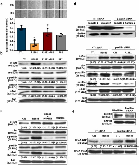
cSrc-mediated signaling pathway is involved in the R1881-induced migration inhibition in HUVECs. (a) Treatment with R1881 (5 nM) 8 h reduced the migration in HUVECs, and this effect was abolished by pre-treatment with 100 nM of PP2, (a cSrc inhibitor). (b) Treatment with R1881 (5 nM) for 2 min increased the levels of p-cSrc, p-paxillin and p-FAK in HUVECs, and these effects were abolished by pre-treatment with 100 nM of PP2. (c) Treatment with R1881 (5 nM) for 5 min increased the levels of p-cSrc, p-paxillin and p-FAK in HUVECs. Pre-treatment with 4 nM of PF573228 (a FAK inhibitor) abolished the R1881-increased the levels of p-FAK and p-paxillin, but not p-cSrc, in HUVECs. (d) Treatment with R1881 (5 nM) for 5 min increased the levels of p-cSrc, p-paxillin and p-FAK in HUVECs. Transfection with paxillin siRNA, which knocked-down the expression of paxillin (top panel), abolished the R1881-increased the levels of p-paxillin, but not p-cSrc and p-FAK, in HUVECs. (e) Treatment with R1881 (5 nM) for 5 min significantly reduced the levels of Rho-GTP in HUVECs. Transfection with paxillin siRNA, which knocked-down paxillin expression (top panel), abolished the R1881-reduced RhoA activity in HUVECs. Values shown in parentheses represent the quantitative results after adjusted with their own input levels and expressed as fold of their own control. (n = 3). *p < 0.05 different from corresponding control. #p < 0.05 different from the R1881-treated group. siRNA, NT-siRNA, non-target siRNA; small interfering RNA. |