
Fig. 1
- ID
- ZDB-FIG-250103-43
- Publication
- Huo et al., 2024 - Androgen receptor activation inhibits endothelial cell migration in vitro and angiogenesis in vivo
- Other Figures
- All Figure Page
- Back to All Figure Page
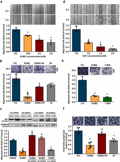
AR activation reduces the migration activity in HUVECs. The cell migration was examined at 9 h after treatment with R1881, DHT or T-BSA using wound healing assay. (a) R1881 (0.05–5.0 nM) concentration-dependently reduced HUVECs migration. Top panel: representative photographs of wound healing migration assay. Bottom panel: quantified results expressed by fold of control. (b) The effect of R1881 on HUVECs transwell migration capacity stained with crystal violet at 24 h (200-fold magnification). The R1881 (5 nM)-reduced endothelial migration was abolished by pre-treatment with HF (5 nM), an AR inhibitor. (c) AR expression was knocked down by AR shRNA (Top panel). The R1881-induced migration inhibition in HUVECs was abolished by knockdown of AR using the shRNA technique. (d) DHT (0.5–2.0 nM) concentration-dependently reduced HUVECs migration. Top pane: representative photographs of wound healing migration assay. Bottom panel: quantified results expressed by fold of control. (e) The effects of R1881 on cell migration detected by transwell assay at 24 h. Treatment with membrane-impermeable T-BSA significantly reduced the migrated cell number in HUVECs. (f) Treatment with R1881 (5 nM) for 4 h reduced the capillary-like tube formation, and this effect was abolished by pre-treatment with HF (5 nM). Values represent the means ± s.e.mean. (n = 3). *p<0.05 different from DMSO-treated group. #p < 0.05 different from the R1881-treated group. DHT, dihydrotestosterone; HF, hydroxyflutamide; shRNA, short hairpin RNA; T-BSA, testosterone-bovine serum albumin. |