
Fig. 3
- ID
- ZDB-FIG-250103-25
- Publication
- Herbert et al., 2024 - Identification of a Specific Granular Marker of Zebrafish Eosinophils Enables Development of New Tools for Their Study
- Other Figures
- All Figure Page
- Back to All Figure Page
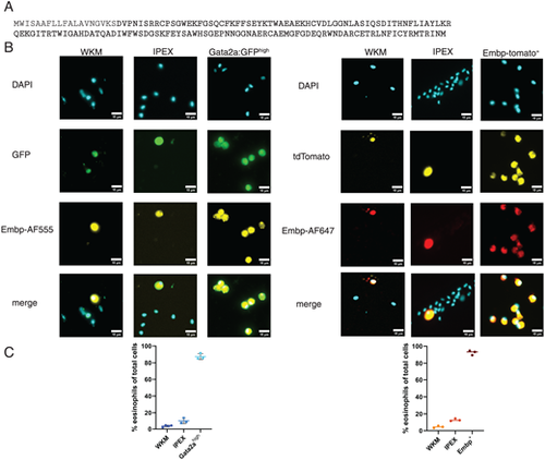
Immunofluorescence staining with a novel polyclonal Ab targeting Embp. (A) Amino acid sequence of Embp in single letter code demonstrating in bold the part of the Ag used to generate the polyclonal Ab. (B) Staining of isolated WKM, IPEX, and eosinophil cell populations of both the Tg(gata2:eGFP) and TgKI(embp-tdTomato,cryaa:EGFP) lines. Eosinophils were sorted by transgenic reporter expression (either Gata2ahigh or Embp+). n = 3. (C) Quantification of cells stained positively for Embp with polyclonal Ab in the aforementioned sorted cell populations. Positive staining was assessed in a minimum of six random fields of view across three independent experiments; 300–500 cells were evaluated per sample. Data are represented as percentage of positive Embp staining in total cell population, including mean with SD of each independent experiment. |