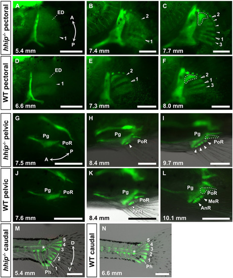
Developmental processes of the fin skeletons in hhip−/−zebrafish. (A-F) Pectoral fin development in hhip−/− (A-C) and WT (D-F) zebrafish. Filled and unfilled white arrowheads indicate new and previous subdivisions, respectively, and numbers mark the predicted first to third subdivisions. White dashed lines indicate the first proximal radial (PR1). (G-L) Pelvic fin development in hhip−/− (G-I) and WT (J-L) zebrafish. White arrowheads indicate the radial bones. AnR, anterior large radial; MeR, medial small radial; Pg, pelvic girdle; PoR, posterior elongated radial. (M,N) Caudal fin development in hhip−/− (M) and WT (N) zebrafish. Numbers indicate the first to fifth hypurals. The white asterisk marks the first ural vertebra. Ph, parhypural. These observations were conducted on more than ten larvae at each developmental stage. Green fluorescence indicates the endochondral skeleton marked by col2a1a:EGFP. Double arrows indicate the anterior (A)-posterior (P) axis and the dorsal (D)-ventral (V) axis. The standard length of individuals (in mm) is shown in the bottom left of each panel. Scale bars: 250 µm.
|
