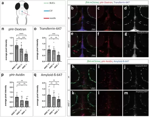
Internalization of different fluorescently labelled substances from the CSF is affected in BLECs devoid of Dab2. (a) Schematic representation of a 5dpf zebrafish embryo from a dorsal view indicating the injection site (red) into the CSF (blue). BLECs are represented in green. The dotted rectangle marks the region of imaging. (b,e and h,k) Maximum projection of a flt4:mCitrine positive wild type sibling (b-d, h-j) or dab2-/- (e-g, k-m) zebrafish embryo injected either with pHr-Dextran and Transferrin-647 (b-g) or with pHr-Avidin and Amyloid-ß-647 (h-m) at 5dpf. Images were taken 1hpi. For each substance, single channels of the respective composite pictures are shown separately. Note the higher signal intensity within BLECs in wild type (c, d, i, j) compared to dab2-/- embryos (f, g, l, m). (n-q) Quantification of pHr-Dextran (n) and Transferrin-647 (o) uptake in BLECs reveals significantly higher average pixel intensities in wild type compared to dab2-/- embryos (ANOVA, t-test; pHr-Dextran: p = 2e-7, Transferrin-647: p = 0.0008). The difference in average pixel intensities for Avidin (p) and Amyloid-ß-647 (q) between wild type and to dab2-/- embryos is not statistically significant (Kruskal Wallis, Mann Whitney U-test; pHr-Avidin: p = 0.09, Amyloid-ß-647: p = 0.296). Graphs show the mean ± SD of 2 (Amyloid-ß) to 3 (Dextran, Transferrin, Avidin) replicates and the individual data point for each embryo (dots). The average pixel intensities are normalized to wild type embryos. CSF, cerebrospinal fluid; dpf, days post fertilization; hpi, hours post injection; pHr, pH-rodo; SD, standard deviation.
|
