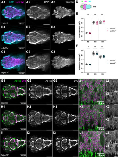
Brain morphology and size is normal in cc2d2a and talpid3 mutants. (A-C) Whole-mount single optical slice widefield immunofluorescence images showing brain morphology of control (A), cc2d2a−/− (B) and talpid3−/− (C) larvae at 5 dpf, stained with anti-HuC/HuD to label neurons (HuC/HuD – magenta) and DAPI to counterstain nuclei (DAPI – cyan). Note that the morphology of the brain in cc2d2a−/− and talpid3−/− larvae is comparable to controls. (D) Schematic showing regions of interest used for brain area quantification. (E,F) Scatter plots showing normalised area of the forebrain, midbrain and hindbrain regions in control versus cc2d2a−/− larvae (E) and control versus talpid3−/− larvae (F) at 5 dpf. There is no statistically significant difference in normalised brain area in cc2d2a−/− larvae compared to controls. In talpid3−/− larvae, there is a minimal but statistically significant difference in normalised forebrain area compared to controls. Each data point represents one larva. Error bars are mean±s.d. ns, not significant; *P≤0.05. Unpaired t-test. For E, control n=22 (N=2), cc2d2a−/− n=14 (N=2) larvae. For F, control n=23 (N=2), talpid3−/− n=22 (N=2) larvae. (G-L) Whole-mount single optical slice widefield immunofluorescence images showing an overview of axonal tracts labelled with anti-acetylated tubulin (AcTub – green) and synaptic neuropil labelled with anti-synaptic vesicle 2 (SV2 – magenta) in control (G, n=35, N=4), cc2d2a−/− (H, n=14, N=2) and talpid3−/− (I, n=24, N=2) larvae at 5 dpf. (J-L) shows higher resolution whole-mount single optical slice confocal immunofluorescence images of boxed region in (G-I). The morphology of axonal tracts and synaptic neuropil appears unchanged in both mutants compared to controls. All images show a dorsal view of 5 dpf larvae with anterior to the left. Scale bars: 500 µm in A-C; 300 µm in G-I and 10 µm in J-L. N denotes the number of independent experiments (i.e. larvae from independent clutches) used for analysis. Analyses were carried out using mz cc2d2a−/− and zygotic talpid3−/− mutants.
|
