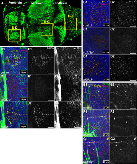
Abnormal cilia in the brain of cc2d2a and talpid3 mutants. (A) Whole-mount confocal image of a 5 dpf zebrafish larva immunostained with anti-acetylated tubulin, providing orientation for the figure. Anterior is to the left and boxes show the analysed brain regions in the subsequent panels. (B-G) Whole-mount maximum projection confocal immunofluorescence images of the forebrain (B-D) and midbrain (E-G), showing primary cilia labelled with anti-Arl13b (Arl13b – yellow) and basal bodies labelled with anti-Centrin (Centrin – magenta) in control (n=12 in B, n=13 in E, N=2), cc2d2a−/− (n=9 in C, n=9 in F, N=1) and talpid3−/− (n=6 in D, n=6 in G, N=2) larvae at 5 dpf. In E-G, Arl13b-positive and acetylated tubulin-positive motile cilia are visible at the midline (indicated with white arrow heads), while asterisks indicate location of Arl13b-positive primary cilia in the midbrain. In both cc2d2a−/− and talpid3−/− larvae, there is a marked reduction in primary cilia. (H-J) Whole-mount maximum projection confocal immunofluorescence images of the hindbrain ventricle, using anti-acetylated tubulin (AcTub – green) and anti-Arl13b (Arl13b – yellow) to label motile cilia and anti-Centrin (Centrin – magenta) to label basal bodies, in control (n=10, N=2), cc2d2a−/− (n=8, N=1) and talpid3−/− (n=6, N=2) larvae at 5 dpf. Motile cilia are reduced in both cc2d2a−/− and talpid3−/− larvae compared to controls. All images show a dorsal view of 5 dpf larvae with anterior to the left. Scale bars: 20 µm. N denotes the number of independent experiments (i.e. larvae from independent clutches) used for analysis. Analyses were carried out using mz cc2d2a−/− and zygotic talpid3−/− mutants.
|
