
Fig. 5
- ID
- ZDB-FIG-241022-31
- Publication
- Singer et al., 2024 - Ultrahigh field diffusion magnetic resonance imaging uncovers intriguing microstructural changes in the adult zebrafish brain caused by Toll-like receptor 2 genomic deletion
- Other Figures
- All Figure Page
- Back to All Figure Page
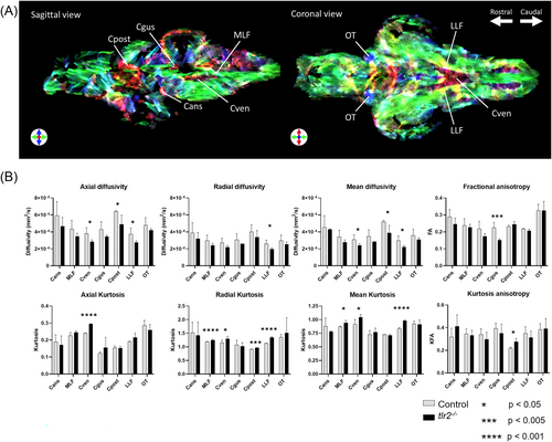
Comparison of DTI results of white matter structures of tlr2−/− and control adult zebrafish. (A) Sagittal and coronal slices of the DEC stTDI msmt-CSD map, used for the identification of white matter structures in the zebrafish brain. Acquisition details: TR 2000 ms, TE 9 ms, 4 averages, isotropic resolution 35 μm, effective b-value range 100, 1000 or 2500 s/mm2, with 4, 12 and 24 directions, respectively. (B) Diffusion metrics estimated by DTI results show reduced D∥, D⊥ and MD, reduced FA, increased K∥, K⊥ and MK and KFA in white matter structures in the tlr2−/− zebrafish brain. Acquisition details: TR 2000 ms, TE 12.4 ms, 32 averages, resolution 25 × 25 × 200 μm, effective b-value range 4, 1000, 3500 or 6000 s/mm2, with 8, 12, 24 or 36 directions, respectively. Statistical analysis was performed using the unpaired t-test, assuming Gaussian distribution, with p less than 0.05 considered to imply significant differences between the control and the tlr2−/− group. *p < 0.05, ***p < 0.005, ****p < 0.001. Cans, ansulate commissure; Cgus, commissure of the secondary gustatory nuclei; Cpost, posterior commissure; CSD, constraint spherical deconvolution; Cven, ventral rhombencephalic commissure; D∥, axial diffusivity; D⊥, radial diffusivity; DEC, directional encoded colour; DTI, diffusion tensor imaging; FA, fractional anisotropy; K∥, axial kurtosis; K⊥, radial kurtosis; KFA, kurtosis fractional anisotropy; LLF, lateral longitudinal fascicle; MD, mean diffusivity; MK, mean kurtosis; MLF, medial longitudinal fascicle; msmt, multishell multitissue; OT, optic tract; stTDI, short-track track-density imaging. |