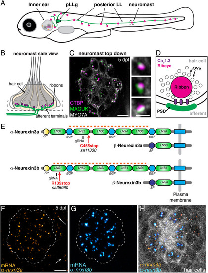
α-nrxn3a and α-nrxn3b are expressed in lateral-line hair cells. (A) Diagram of a 5 day post fertilization (dpf) larval zebrafish showing sensory hair-cell clusters in the inner ear and posterior lateral line (neuromasts). Neurons from the posterior lateral line ganglia (pLLg) innervate (pLLg, green) lateral-line neuromasts. (B) Side view of a neuromast organ showing hair cells, presynapses (ribbons) and afferent processes. The dashed box outlines the synaptic layer. (C) Immunostaining of the synaptic layer, viewed from above. CTBP and MAGUK stain presynapses/ribbons (magenta) and postsynapses (green), respectively. MYO7A stains hair cells (gray). Higher magnification of three synapses is shown on the right. (D) Diagram of a hair-cell ribbon synapse. The presynapse/ribbon consists mainly of Ribeye, a splice variant of CTBP2. The ribbon is surrounded by glutamate-filled synaptic vesicles (SVs). CaV1.3 channels cluster beneath the ribbon, opposite the postsynaptic density (PSD). (E) Zebrafish have two orthologues of Nrxn3, Nrxn3a and Nrxn3b. Each neurexin has a long α form and a shorter β form. Red arrows show location of the mutations in germline zebrafish mutants; these lesions disrupt an obligatory exon in the α form of each orthologue (C455stop and R134stop). Location of gRNAs used in a crispant analysis are indicated. The α and β forms each have a unique start and signal peptide (SP). Each α form has six Laminin G-like domains (LNS) and three epidermal growth factor-like domains (EGF). The red dashed line indicates the location of the RNA FISH probes used in F-H. (F-H) RNA FISH analysis reveals that both α-nrxn3a (F, orange) and α-nrxn3b (G, cyan) mRNAs are present in lateral-line hair cells. Arrows in F indicate α-nrxn3a puncta that are present outside of hair cells, likely in supporting cells. In H, hair cells (myo6b:memGCaMP6 s) are shown in grayscale. The dashed lines in F-H outline the location of hair cells. All images are from larvae at 5 dpf. Scale bars: 5 µm (C,F): 1 µm (C, inset).
|

