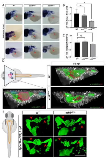
Ezh2 mediates isl1 regulation with tissue specificity on the nephric region and causes defective nephric duct development. (A) isl1 in situ hybridization in WT, ezh2 +/- and KO larvae at 56 hpf. Red arrows indiacate the expression of isl1 in the nephric region that shows clear staining in the WT and ezh2 +/- lines and a strong reduction in the ezh2 KO zfl. Isl1 expression results almost not alterated in the brain and spinal chord of all genotypes. (B) Normalized isl1 qPCR in head-chopped embryo at 56 hpf shows a significantly reduced signal in the ezh2 KO line. (C) Normalized isl1 qPCR of whole zfl at 56 hpf shows no significant reduction of isl1 expression. (D) Isl1 immuno histochemistry (red cells) in Tg(wt1b: eGFP) line (in green) and double transgenic Tg(wt1b: eGFP) – ezh2 KO line. Left pannel indicates the location of the transversal paraffin section in reference of the whole zfl and nephric region. Blue circles indicate the sagittal nephric ducts; purple circle indicates the glomeruli region, white asterics the sagittal nephric ducts and the white plus the pancreas. Right pannels show the 3D co-localization of Isl1 protein (red) on the glomeruli and nephric ducts (green) in the WT (top) and ezh2 KO (lower) 56 hpf zfl. A clear absence of Isl1 signal locates to the glomeruli and nephric ducts of the ezh2 KO line compared to WT. (E) Nephric ducts of the ezh2 KO larvae display developmental defects and malformation at 3 dpf. Red arrows indicate the correct protrusion of the nephric duct in the WT and absence of GFP signal, togeather with dilated nephric ducts in the ezh2 KO.
|
