|
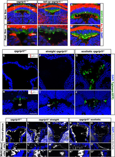
Presence of scospondin aggregates and heterogenous ciliary defects in the fChP of rpgrip1l-/- juvenile fish. (A-B’) Immunostaining on transverse sections of the SCO, at anterior level (A, A’, B) or posterior level (C, D) in rpgrip1l+/+ (n=3) and tail-up rpgrip1l-/- (n=3), transgenic for Sspo-GFP (N=2). Sspo-GFP forms small cytoplasmic aggregates within SCO cells of both genotypes (* in A’ and B’), under the primary cilia extending towards the DiV, and accumulates at the apical surface of tail-up rpgrip1l-/-, creating a double positive area for cilia and Sspo-GFP staining (yellow colour in B’). Multiciliated cells lateral to the posterior SCO (mcc) are negative for Sspo-GFP staining in controls (C) and absent in tail-up rpgrip1l-/- (D). (E–J) GFP immunostaining on transverse section of the fChP (E–G) or at the level of the optic tectum (H–J) in rpgrip1l+/+ (n=7), straight rpgrip1l-/- (n=4) and scoliotic rpgrip1l-/- (n=5) 8 wpf fish transgenic for sspo-GFP (N=2). Scoliotic rpgrip1l-/- fish present with filamentous material and aggregates within the ventricles (G, J) while a small RF fragment is present within the ventricle of controls (H) and straight mutants (I) at optic tectum level. * indicates aggregates. (K-O’’) Representative maximum intensity Z-stack projections of confocal transverse brain section at the level of the fChP in rpgrip1l+/+ (K-K’’) (n=7), straight rpgrip1l-/- (L-M’’) (n=4) and scoliotic rpgrip1l-/- (N-O’’) (n=5) 8 wpf fish (N=2). Cilia were stained with glutamylated-tubulin and acetylated-tubulin antibodies (both in white) and nuclei were stained with DAPI (blue). Sections shown are at the level of the habenular nuclei. At this level, monocilia in the dorsal midline region (K’) and multicilia on lateral sides (K’’) (n=7/7) are present in controls. Straight rpgrip1l-/- exhibited either normal (L’, L’’) (n=2/4) or abnormal and sparser (M’, M’’) (n=2/4) monocilia and multicilia. Similarly, scoliotic rpgrip1l-/- presented either normal monocilia and multicilia (N’, N’’) (n=3/5) or long monocilia (O’) and decreased cilia density of multi-ciliated tufts (O’’) (n=2/5). Scale bars: 20 µm.
|