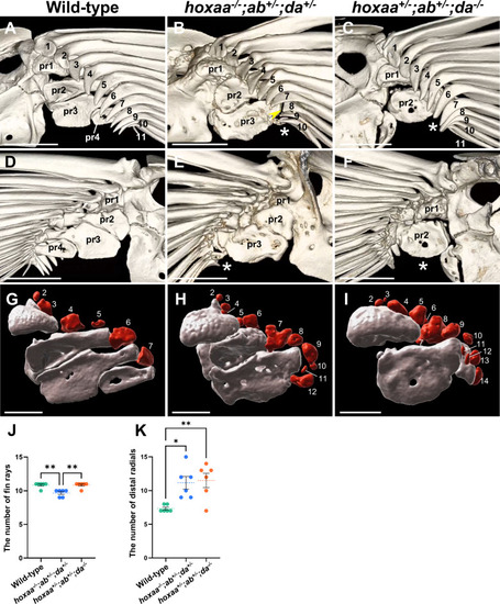
Skeletal elements in the posterior regions of the pectoral fin are preferentially affected in hoxaa−/−;hoxab+/−;hoxda+/− and hoxaa+/−;hoxab+/−;hoxda−/− adult fish. Micro-CT scan analysis of the skeletal structures in zebrafish pectoral fins. Both sides of the pectoral fins in wild-type (n = 3), hoxaa−/−;hoxab+/−;hoxda+/− (n = 3), and hoxaa+/−;hoxab+/−;hoxda−/− (n = 3) adult zebrafish were analyzed. The left pectoral fins are shown. (A–C) The magnified views of pectoral fin endoskeletons from the lateral side. (D–F) Magnified views of the pectoral fin endoskeletons from the opposite medial side in the same specimens shown in A-C. Proximal radials (pr1-pr4) are indicated. Fin rays are numbered from the anterior to the posterior. The asterisk indicates the absence of the posterior proximal radials. The arrowhead indicates the loss of the proximal tip of the fin rays. Anterior is to the top. The 3D movies were created using OsiriX MD ver13 and are available in Movies S1–3. Scale bar, 500 μm. (G–I) Magnified images of proximal radials (grey) and distal radials (red) as revealed by CT segmentation. The same specimens as above were used for each genotype. Fin rays were removed by image processing. The first distal radials are not shown due to their distance from the remaining distal radials. 3D movies were created using Imaris ver10 (Movies S4–6). A 2D movie showing the distal radials and proximal radials was also created using Imaris ver10 and is provided in Movie S7. Scale bar, 250 μm. (J, K) Comparisons of the number of fin rays and distal radials between wild-type, hoxaa−/−;hoxab+/−;hoxda+/− and hoxaa+/−;hoxab+/−;hoxda−/− adult fish. The Tukey-Kramer test was performed with *P < 0.05 and **P < 0.01.
|
