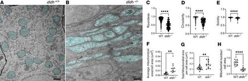
Intestinal mitochondria in 7 dpf dldh–/– larvae showed depletion of mitochondria with matrix chamber swelling and elongation. (A and B) Mitochondria ultrastructure (light blue) was nicely rounded in WT intestinal cells with well-organized rough reticulum (A), while intestinal cells of 7 dpf dldh–/– zebrafish larvae showed mitochondria (light blue) swelling, damage, and elongation (B). (C–E) Mitochondria ultrastructural morphological features quantified in intestinal cells of WT and dldh–/– larvae mutant larvae. Mitochondria, n = 504 (WT) and 190 (dldh–/–) in 16 cells (WT) and 13 cells (dldh–/–) from 3 WT and 3 dldh–/– larvae. (F–H) Average mitochondrial area, total mitochondrial area, and mitochondria number relative to cross-sectional area . Analysis confirmed increased mitochondrial area and decreased number of mitochondria in dldh–/– larvae relative to WT. All characteristics determined with ImageJ; statistical analyses by Student’s t test, and each point represents data from 1 cell. ****P < 0.0001; **P < 0.01. Data are shown as mean ± SD.
|
