
Fig. 1
- ID
- ZDB-FIG-240918-106
- Publication
- Giese et al., 2024 - Staphylococcus aureus lipid factors modulate melanoma cell clustering and invasion
- Other Figures
- All Figure Page
- Back to All Figure Page
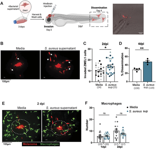
S. aureus supernatant promotes melanoma invasion and dissemination in vivo. (A) Schematic of ZMEL1-td-Tomato melanoma cell injection into larval zebrafish hindbrain, created with BioRender.com. Representative image of ZMEL1 dissemination to tail fin of 4dpi zebrafish larvae. Confocal imaging was performed in the hindbrain at 2 days post injection (dpi) and in the tail at 4 dpi. (B,C) Representative images of ZMEL1 melanoma cell invasion (B) and quantification of ZMEL1 melanoma cell invasion (C) at 2 dpi in the zebrafish hindbrain after culture in medium alone (Media) or in S. aureus USA300 supernatant. Arrows in B indicate migrated melanoma cells with an elongated morphology. Results are from three independent experiments (n=24-25 larvae per condition). Dots in C represent independent larvae color coded per replicate. (D) Percent of zebrafish with disseminated ZMEL1 melanoma cells in the tailfin at 4 dpi. Results are from three independent experiments (n=130 larvae for medium control, n=119 larvae with S. aureus supernatant). Dots represent independent replicates. Error bars indicate the mean±s.e.m. (E,F) Representative images of macrophage recruitment to ZMEL1 melanoma cells (E) and quantification of macrophage recruitment to ZMEL1 melanoma cells (F) at 2 dpi. Areas surrounded by dashed line indicate the 50-μm region within which recruited immune cells were counted Results are from two independent experiments (n=17-20 larvae per condition). Error bars indicate the mean±s.e.m.. P-values were calculated using unpaired t-test (C), paired t-test (D) or two-way ANOVA (F). *P<0.05; **P<0.01, ns, not significant. |