
Fig. 2
- ID
- ZDB-FIG-240829-96
- Publication
- Han et al., 2024 - Evaluation of the safety and efficacy of cosmetics ingredient Spherulites Paeony Superior Retinol
- Other Figures
- All Figure Page
- Back to All Figure Page
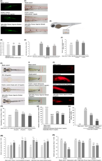
The skin care efficacies of Spherulites Paenoy Superior Retinol were investigated using zebrafish embryos. Spherulites Paenoy Superior Retinol of 5 μg/mL was used in all seven experiments. (A, B) The anti-ROS experiment exposed 48 hpf embryos to Spherulites Paenoy Superior Retinol solution for 24 h, H2DCFDA stained with fluorescence signal intensity measured. The positive control 0.1 mg/mL glutathione reduced 13% (p = 0.0011) and Spherulites Paenoy Superior Retinol reduced 7.4% (p = 0.028) ROS compared with the blank control. (C, D) The anti-inflammation experiment treated 72 hpf zebrafish embryos with Spherulites Paenoy Superior Retinol solution in the presence of 10 μM CuSO4 (inflammation inducer) for 40 min, Sudan Black B stained, with neutrophils (stained brown dots) in the lateral midline region (red dash line circled) counted. The positive control 10 μM indomethacin reduced 30% (p < 0.001) and Spherulites Paenoy Superior Retinol reduced 20% (p < 0.001) neutrophil aggregation compared with the model control. (E, F) The blood circulation experiment exposed 72 hpf zebrafish embryos to Spherulites Paenoy Superior Retinol solution for 2 h, with dorsal aorta blood circulation speed measured. The positive control 50 μg/mL caffeine increased 28% (p < 0.001) and Spherulites Paenoy Superior Retinol increased 14% (p = 0.0063) blood circulation speed compared with the blank control. (G, H) The whitening experiment exposed 6 ~ 8 hpf zebrafish embryos to Spherulites Paenoy Superior Retinol solution till 48 hpf with embryo transmittance signal measured to correlate melanin level. The 30 μg/mL PTU inhibited embryos melanin development completely (100% whitening efficacy, p < 0.001), positive control 2.5 mg/mL kojic acid reduced 45% (p < 0.001) and Spherulites Paenoy Superior Retinol reduced 10% (p < 0.001) melanin development. (I, J) The wound-healing experiment exposed tail fin amputated zebrafish embryos to Spherulites Paenoy Superior Retinolsolution for 48 h, with tail fin length measured. The positive control 0.1% Rehmannia glutinousa Libosch extract enhanced 31% (p < 0.001) and Spherulites Paenoy Superior Retinol enhanced 9.0% (p = 0.030) tail fin regeneration. (K, L) The skin barrier protection experiment exposed 72 hpf zebrafish embryos to Spherulites Paenoy Superior Retinol solution for 4 h with the 1.1 mg/mL lactic acid and 0.02 mg/mL crystal violet were added to the solution for 20 min, using fluorescence area in the embryo tail region as the measurement. The positive control 150 μg/mL ectoin reduced 33% (p < 0.001) and Spherulites Paenoy Superior Retinol reduced 72% (p < 0.001) crystal violet penetrating through the skin barrier. (M, N) The gene regulation experiment exposed 4 dpf zebrafish embryos to Spherulites Paenoy Superior Retinol solution for 24 h, and the relative gene expression level by qRT-PCR was analyzed using β-Actin as the house-keeping gene. Type I collagen genes (col1a1a, col1a1b and col1a2) and elastin gene (elna) expression analysis showed that both positive control acetyl hexapeptide-8 (0.8 mg/mL) and Spherulites Paenoy Superior Retinol significantly (p < 0.05) increased the expression of these genes, and 5α-reductase genes (czsrd5a1, zsrd5a2a and zsrd5a2b) expression analysis revealed that positive control dutasteride (1 μM) and Spherulites Paenoy Superior Retinol significantly (p < 0.05) reduced the expression of these genes. Data was presented as mean ± SEM. “###” denotes compared with blank control, p < 0.001. Compared with blank control or model wherever model included, “*” p < 0.05, “**” p < 0.01, “***” p < 0.001. |