Fig. 8
|
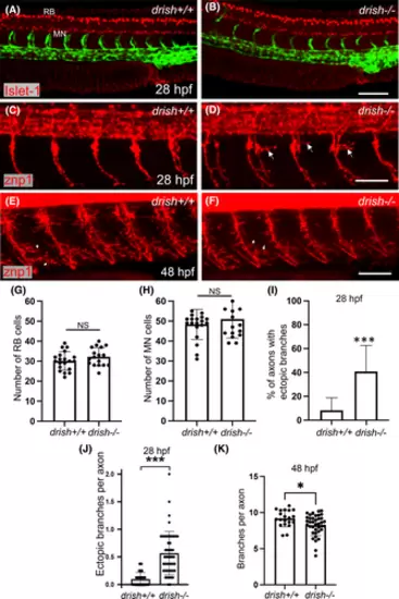
Analysis of Rohon-Beard (RB) neurons and spinal motor neurons (MNs) in drish mutants. (A, B) Confocal imaging of drish+/+ and drish−/− embryos in Tg(fli1a:GFP) background processed for Islet-1 immunostaining at 28 hpf, which labels RB neurons and spinal MNs. Lateral view of trunk region with anterior to the left shown. Scale bar, 100 μm. (C–F) Immunostaining for Znp-1 at 28 and 48 hpf, which labels primary motor neurons. drish−/− mutants exhibited ectopic axon branching of caudal primary motor neurons and the formation of peripheral branches at 28 hpf (arrows, D). In contrast, the number of peripheral branches of CaP neurons was reduced at 48 hpf (arrows, E, F). Scale bar, 50 μm. (G, H) Quantification of RB neurons and spinal MNs at 28 hpf. n = 20 for drish+/+ and n = 17 for drish−/− embryos. No difference in the number of RB neurons (average number of RB neurons in drish+/+: 30.2 and drish−/−: 32.2, p = 0.17, Student's t-test) and spinal MNs (average number of spinal MNs in drish+/+: 48.4 and drish−/−: 51, p = 0.34, Student's t-test) was observed between drish+/+ and drish−/− embryos. NS, not significant. (I–K) Quantification of axon branching defects in drish−/− mutants at 28 and 48 hpf. Percentage of axons with ectopic branches in each embryo and the number of ectopic branches per axon at 28 hpf are shown in (I, J), while the total number of peripheral branches per ventral segment of an axon at 48 hpf is shown in (K). n = 35 for drish+/+ and n = 48 for drish−/− at 28 hpf (I, J); n = 20 and 40 for drish+/+ and −/− embryos, respectively, at 48 hpf (K). The number of embryos analyzed was combined from two independent experiments. ***p < 0.0001, *p < 0.05, Student's two-tailed t-test (J, K), Fisher's exact test performed using absolute numbers of axon counts (I). |

