|
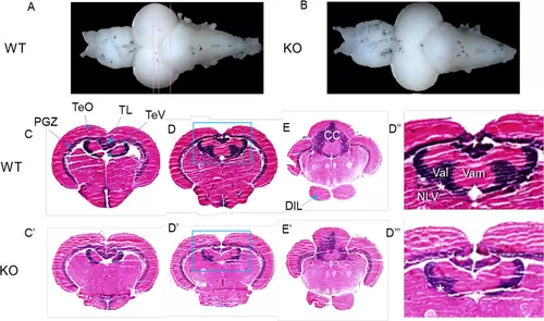
Reduced size of the valvular cerebelli in adult brain of srpk3 KO zebrafish. (A, B) Representative whole brains dissected from a control wild-type (WT) and a knockout (KO) zebrafish. Doral view, anterior is to the left. (C, D‴) Anatomical analysis of brain sections after H&E stain. Frontal sections at 3 different levels indicated in (A). (D″–D‴) Enlargement of valvular cerebelli region in the inlets in (D) and (D′). Asterisk indicates a structural connection between the nucleus lateralis valvulae (NLV) and the valvular cerebelli. Number of adult zebrafish brain used for analysis; n = 3 for WT and n = 3 for KO. CC = corpus cerebelli; DIL = diffuse nucleus of the inferior lobe; NLV = nucleus lateralis valvulae; PGZ = periventricular gray zone of optic tectum; TeO = tectum opticum; TeV = tectal ventricle; TL = torus longitudinalis; Val = lateral division of valvular cerebelli; Vam = medial division of valvular cerebelli.
|