
Fig. 3
- ID
- ZDB-FIG-240729-146
- Publication
- Bludau et al., 2024 - Inflammation is a critical factor for successful regeneration of the adult zebrafish retina in response to diffuse light lesion
- Other Figures
- All Figure Page
- Back to All Figure Page
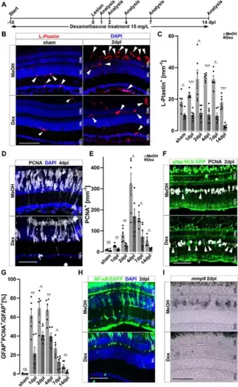
Immunosuppression interferes with leukocyte accumulation and Müller glia reactivity. (A) Scheme of experimental outline. Fish were treated with Dexamethasone (Dex) or vehicle (Methanol; MeOH) from 10 days prior to injury until the day of analysis (sham, 1, 2, 4, 7 and 14 days post lesion; dpl). (B) Dex-treatment reduces the number of L-Plastin+ leukocytes (arrowheads) in sham (left panel) and regenerating retinae at 2 dpl (right panel). (C) Quantification of L-Plastin + cells in MeOH- and Dex-treated sham or lesioned animals at 1, 2, 4, 7 and 14 dpl. (D) Immunohistochemistry for proliferating cell nuclear antigen (PCNA) reveals impaired proliferation in Dex-treated animals at 4 dpl. (E) Quantification of PCNA + cells in MeOH- and Dex-treated sham and regenerating retinae at 1, 2, 4, 7 and 14 dpl indicates impaired proliferative response in the Dex-treated group. (F) Immunohistochemistry for PCNA in gfap:NLS-GFP labeled Müller glia shows reduced numbers of proliferating Müller glia in the Dex treated group at 2 dpl compared to MeOH controls (arrowheads indicating non proliferative Müller cells). (G) Quantification of proliferating Müller glia in vehicle and Dex-treated sham and regenerating retinae at 1, 2, 4, 7, and 14 dpl, indicating that Dex hinders Müller glia proliferation. (H) In comparison to vehicle, the NFκB:GFP reporter is not activated in the inner nuclear layer (INL) of Dex-treated retinae at 2 dpl. (I) Injury-induced expression of mmp9 is strongly reduced in Dex-treated retinae at 2 dpl. Retinal layering is indicated by dashed lines. Scale bar: 50 μm, Error bars indicate SEM; ns = p > 0.05, * = p ≤ 0.05; ** = p ≤ 0.01; *** = p < 0.001; N = 6; two-tailed t-Test, ONL = outer nuclear layer, INL = inner nuclear layer GCL = ganglion cell layer. |