
Fig. 1
- ID
- ZDB-FIG-240729-144
- Publication
- Bludau et al., 2024 - Inflammation is a critical factor for successful regeneration of the adult zebrafish retina in response to diffuse light lesion
- Other Figures
- All Figure Page
- Back to All Figure Page
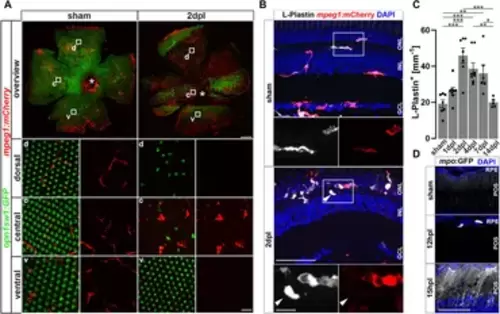
Sterile phototoxic ablation of photoreceptors triggers leukocyte accumulation. (A) Retinal flat mounts of Tg(opn1sw1:GFP) x Tg(mpeg1:mCherry) show the ramified structure of leukocytes in a UV-cone-specific reporter line and their reaction to lesion. Upon light lesion, leukocytes increase in number at the lesion site and display an amoeboid, activated morphology and accumulate in the central part of the lesion, while unharmed areas appear devoid of mpeg1:mCherry positive cells. A central stripe of autofluorescence can be seen (dashed lines) at 2 dpl. In the corresponding inset a few remaining UV-cones are visible. The optic nerve head is indicated by an asterisk. (B) Retinal sections show that the majority of L-Plastin+ cells are Tg(mpeg1:mCherry)+, display morphological changes after lesion, and accumulate at the outer nuclear layer (ONL) in response to lesion. However, L-Plastin+ Tg(mpeg1:mCherry) negative cells were also observed upon injury (arrowhead). (C) Quantification on sections of L-Plastin+ cells in sham and regenerating retinae at 1, 2, 4, 7 and 14 days post lesion (dpl) shows an increase of cells within 2 dpl that is resolved within 14 dpl. (D) In contrast to sham, Tg(mpo:GFP)+ neutrophils are detected upon lesion. They gather at the lesion site (12 hpl) and a diffuse GFP positive pattern (matrix) is formed from 15hpl onwards. Scale bars in A: overview 200 μm, Insets 10 µm; Scale Bars in D: overview 50µm, insets 10µm; error bars indicate SEM; * = p ≤ 0.05; ** = p ≤ 0.01; *** = p < 0.001; N ≥ 6; one-way ANOVA Tukey’s post hoc analysis; ONL = outer nuclear layer, INL = inner nuclear layer; GCL = ganglion cell layer; POS = photoreceptor outer segments; RPE = retina pigment epithelium; d = dorsal; c = central; v = ventral. |