
Fig. 3
- ID
- ZDB-FIG-240725-30
- Publication
- Li et al., 2024 - Biosynthetic deficiency of docosahexaenoic acid causes nonalcoholic fatty liver disease and ferroptosis-mediated hepatocyte injury
- Other Figures
- All Figure Page
- Back to All Figure Page
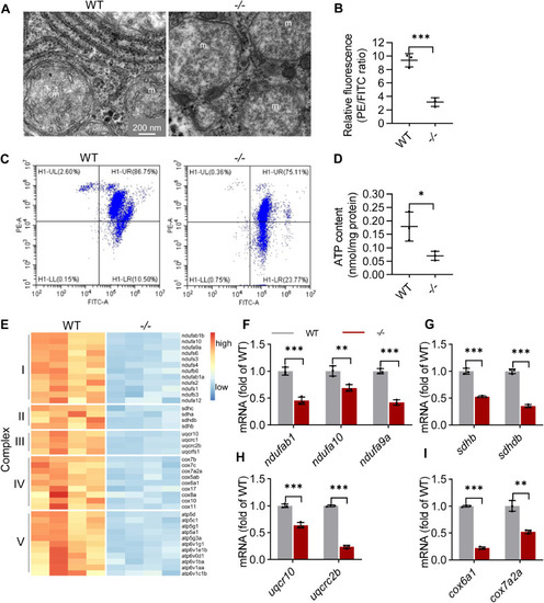
The function of mitochondria was impaired in the elovl2−/− hepatocyte. A, mitochondria ultrastructure in the hepatocytes of WT and −/−. m: mitochondria. Scale bar: 200 nm. B and C, flow cytometric analysis of mitochondrial transmembrane potential after staining with JC-1 and statistical analysis of the PE/FITC fluorescence ratios. D, analysis of ATP production in WT and −/− livers. E, heatmap of genes related with multiheteromeric enzyme complexes of oxidative phosphorylation in WT and −/− livers. F–I, qRT-PCR analysis of genes related with multiheteromeric enzyme complexes of oxidative phosphorylation in WT and −/− livers. N = 3 replicates. All values are mean ± SD. A Student t test was used. ∗p < 0.05, ∗∗p < 0.01, ∗∗∗p < 0.001. Individual p values are listed in Table S5 . −/−, elovl2−/−; WT, wildtype. |