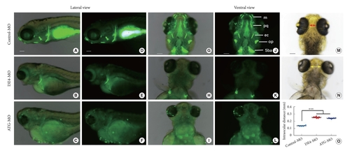
Loss of best1 causes abnormal craniofacial structure and interorbital distance. (A–N) Representative bright field and fluorescent images of zebrafish head skeleton at 6-dpf. In vivo visualization of the skeleton is achieved by the administration of a fluorescent dye (Calcein) directly to the fish water. Dyes that bind to calcified matrix can be used to label the entire skeleton. Lateral view (A–F) and ventral view (G–L) of the head skeleton of day-6 embryos labeled with Calcein. (B, C, E, F, H, I, K, and L) When embryos were injected with best1-morpholino (MO) at the one-cell stage, the amount of stained mineralized tissue was significantly reduced compared to fish injected with control-MO. Panel N show increased intraocular distance (yellow arrows) in best1 morphants. Panel M and N show measurements of the distance between the eyes, and panel O shows the distances depicted graphically as the mean for 10 embryos of each type. Columns, mean; standard error of the mean (n = 10; analysis of variance), ***p < 0.0001. Scale bar, 100 μm. 5ba, fifth branchial arch; op, opercular bone; ec, ectopterygoid; e, ethmoid plate; pq, palatoquadrate; m, Meckel cartilage; dpf, days postfertilization.
|
