
Fig. 3
- ID
- ZDB-FIG-240620-287
- Publication
- Gauvrit et al., 2024 - A β-catenin chromobody-based probe highlights endothelial maturation during vascular morphogenesis in vivo
- Other Figures
- All Figure Page
- Back to All Figure Page
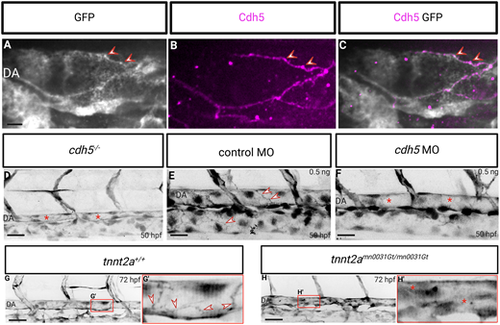
Membrane localization of the endothelial-specific β-catenin chromobody is dependent on Cdh5 expression and blood flow. (A-C) Confocal images of immunostaining (GFP, white; Cdh5, magenta) of Tg(fli1a:EGFP-BC1) embryos at 48 hpf. Cdh5 co-localizes with the chromobody at the endothelial cell-cell junctions (red arrowheads). (D) Trunk vasculature of Tg(fli1a:EGFP-BC1); cdh5−/− embryos at 50 hpf. cdh5 mutants mostly lack blood flow and display a lack of localization of the β-catenin chromobody at endothelial cell-cell junctions (asterisks indicate a collapsed dorsal aorta due to the lack of blood flow). (E,F) Trunk vasculature of 50 hpf Tg(fli1a:EGFP-BC1) embryos injected at the one-cell stage with 0.5 ng control MO (E) or 0.5 ng cdh5 MO (F). cdh5 morphants display a lack of localization of the β-catenin chromobody at endothelial cell-cell junctions while blood flow is not affected (arrowheads point to β-catenin chromobody localization at endothelial cell-cell junctions; asterisks indicate a lack of localization of the β-catenin chromobody at endothelial cell-cell junctions). (G,H) Trunk vasculature of Tg(fli1a:EGFP-BC1); tnnt2a+/+ and Tg(fli1a:EGFP-BC1); tnnt2a−/− (tnnt2amn0031Gt/mn0031Gt) larvae at 72 hpf. tnnt2a mutants display a lack of blood flow, and the localization of the β-catenin chromobody at endothelial cell-cell junctions is missing. (G′,H′) High-magnification images of boxed areas in G and H, showing the dorsal aorta. Arrowheads point to β-catenin chromobody localization at endothelial cell-cell junctions; asterisks indicate a lack of localization of the β-catenin chromobody at endothelial cell-cell junctions. Scale bars: 40 μm. |