
Fig. 2
- ID
- ZDB-FIG-240620-286
- Publication
- Gauvrit et al., 2024 - A β-catenin chromobody-based probe highlights endothelial maturation during vascular morphogenesis in vivo
- Other Figures
- All Figure Page
- Back to All Figure Page
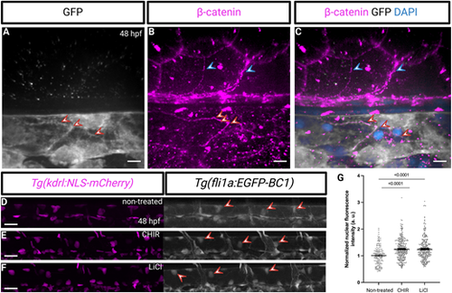
β-Catenin chromobody expression in endothelial nuclei. (A-C) Confocal images of immunostaining (GFP, white; β-catenin, magenta) and counterstaining for DNA (DAPI, blue) of Tg(fli1a:EGFP-BC1) embryos at 48 hpf. β-Catenin immunostaining coincides with the chromobody localization at the endothelial cell-cell junctions (red arrowheads point to endothelial cell-cell junctions, blue arrowheads to non-endothelial β-catenin immunostaining). (D-G) Lateral views and quantification of fluorescence intensity of the nuclear signal in dorsal aorta endothelial cells of 48 hpf Tg(fli1a:EGFP-BC1); Tg(kdrl:NLS-mCherry) embryos after exposure to the indicated GSK3β inhibitor starting at 24 hpf (non-treated, n=166 nuclei from six embryos; CHIR, n=305 nuclei from 12 embryos; LiCl, n=311 nuclei from 12 embryos). Treatment with either GSK3β inhibitor increases the nuclear signal of the Tg(fli1a:EGFP-BC1) chromobody. Data are mean±s.d. P<0.0001 calculated using a one-way ANOVA (post hoc Tukey's test). Red arrowheads point to endothelial nuclei. Scale bars: 40 μm (A-C), 10 μm (D-F). |