
Figure 3
- ID
- ZDB-FIG-240614-72
- Publication
- Abu Nahia et al., 2024 - scRNA-seq reveals the diversity of the developing cardiac cell lineage and molecular players in heart rhythm regulation
- Other Figures
- All Figure Page
- Back to All Figure Page
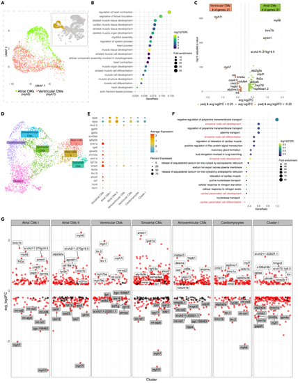
Analysis of the “Myocardium” cluster revealed the diversity of myocardial cells and pinpointed sinoatrial cardiomyocytes within the atrial myocardium (A) Two distinct identities of chamber myocardium could be delineated by molecular markers: Atrial CMs by expression of (B) Gene Ontology enrichment analysis of all genes enriched within the “Myocardium” cluster. (C) Volcano plot depicting differentially expressed genes between atrial and ventricular CMs fractions. (D) UMAP projection of re-clustered myocardial cells reflecting the heterogeneity of atrial and ventricular myocardium. (E) Dotplot showing the expression (SCT normalized counts) of well-established gene signatures associated with working myocardium and sinoatrial pacemaker within “Myocardium” clusters. (F) Gene Ontology enrichment analysis of genes enriched within the “Sinoatrial CMs” subcluster. (G) Differentially expressed genes in each myocardial subclusters. Top 8 genes in terms of significance were labeled. Genes with adjusted |